Day 2 :
- Track 3: Pediatric Dentistry
Track 8: Periodontics
Track 9: Endodontics
Track 10: Future Trends in Dentistry
Track 11: Oral Medicine Diagnosis and Radiology
Track 13: Dental Public Health
Track 14: Regulatory and Ethical Issues in Dentistry
Location: Al Dhiyafah 5-6

Chair
Anka Letic
Rakcods, UAE

Co-Chair
Zikra A Alkhayal
King Faisal Specialist Hospital and Research Center, Saudi Arabia
Session Introduction
Zikra A Alkhayal
King Faisal Specialist Hospital and Research Center, Saudi Arabia
Title: Inhalational Sedation with Nitrous Oxide In Current Pediatric Practice
Time : 11:00-11:20

Biography:
Dr Zikra Alkhayal has completed her undergraduate dental training from the Royal London, England, postgraduate training in Pediatric Dentistry from University of Illinois, Chicago, USA and was the first Saudi to Obtain the American Board of Pediatric Dentistry. Currently, Consultant Pediatric Dentist and Joint Appointee/Scientist, Stem Cell and Regeneration Program in King Faisal Specialist Hospital & Research Centre, Riyadh, Saudi Arabia. She was recently a Visiting Research Fellow, King's College, London, UK and Visiting Research Associate, Harvard School of Dental Medicine, USA. Her current interests are in the development of Special Care Dentistry for the medically compromised,procedural sedation, quality of care and the translational aspects of stem cell and regeneration.
Abstract:
Children often have caries and need dental treatment for their primary, mixed, or permanent dentitions. The ability of children to accept and cope with dental procedures varies along a continuum of cooperativeness and depends on a host of prominent factors including age, cognitive and emotional attributes, personality and temperament characteristics, parental, sibling or peer influences, social skills, degree of pain tolerance, and past experiences in medical, dental or other setting. Most children are relatively easy to treat in the dental operatory and respond well to guidance and information given to them by the dental team. They have coping skills and temperaments that contain any arising anxieties associated with dental procedures. A minority of children are not cooperative. They may have normal cognitive function but poor coping skills for handling situational anxieties and fears associated with dentistry. Others may have emotional, physical or cognitive problems that with poor coping skills cause them to respond in a disruptive and uncontrolled ways.It is often quoted that the majority of children will accept dentistry if managed with routine behavioral techniques but some children primarily due to variably- expressed degrees of fear and/or anxiety, will require more rigorous methods such as sedation to render quality care. Conscious sedation is used in pediatric dentistry as elsewhere to reduce fear and anxiety in pediatric patients and so promote favorable treatment outcomes. This can help to develop a long-term positive psychological response to necessary dental procedures. Currently, different forms of sedation, for example, oral, intravenous, inhalation, intranasal and combinations of treatments are used for pediatric dental patients worldwide. Currently, inhalation sedation using nitrous oxide is suggested as the treatment of choice for pediatric patients in the primary dental care setting. The aim of this presentation is to give an overview of Nitrous Oxide Inhalational Sedation in children and its effectiveness. Further, a discussion of alternative methods of sedation in children will be reviewed to examine the use of Nitrous Oxide Inhalational Sedation in current Pediatric Practice.
Javad Faryabi
Kerman University of medical Sciences, Kerman, IRAN
Title: ÙFacial swellingÙ: sometimes is challenging for dentists
Time : 11:20-11:40
Biography:
Javad Faryabi has graduated from dentistry school of Shiraz University of Medical Sciences in IRAN (1990) and postdoctoral studies from Shahid Beheshti University of Medical sciences in Tehran, IRAN(2000) . He is the director of residency program of maxillofacial surgery in Kerman University,and head of Oral & Maxillofacial deprtment of this university. He has published more than 20 papers in reputed journals and has been serving as an editorial board member of JOHK inthe mentioned above unhversity.
Abstract:
Currently, when a dentist seeks a treatment for facial swelling of his/her patients, the first diagnosis and cause of swelling that he/shet thinks about it, may be and perhaps always is the teeth of the patients and an finally is an odontogenic infection. But sometimes it is not true, and this is not the correct answer to the patients for managing their discomfort, because they are worrying about their health, and may go to several offices of dentists or other physicians and specialists for treatment of their disease, actually they need to additional work ups like CT-Scans, CBCT, MRI, Sonography, and etc.to discover the exact cause of their facial swelling, so In my presentation I will introduce to several example of such facial swelling in my patients related to maxillofacial region and proper management of them.
Gianni Frisardi
Orofacial Pain Centre, Italy
Title: NGF-Neuro Gnathological Functions: A new Trigeminal Electrophysiological Paradigm in masticatory rehabilitation
Time : 11:40-12:00
Biography:
Gianni Frisardi has completed his bachelor’s degree in Medicine and Dentistry in 1983 and 1987 respectively at the “La Sapienza†University in Rome. In 1993 he attained a Master in Bioengineering and his experimental thesis “Mandibular Kinematics has been awarded by the ENEA Institute. He worked for several years in the trigeminal neurophysiology laboratory in order to transfer the know how in the field of the gnathology. He published papers in reputed journals and in 2014 was candidated as Editor by the World Dental Federation for the International Dental Journal. He has been Visiting Professor at the University of Rome, Chieti, Ancona and currently at the University of Sassari in the Department of Masticatory Rehabilitation and Orofacial Pain.
Abstract:
The masticatory system should be considered a "Complex System", which is divided into a number of constituent elements bound toghether in a set of non-linear and stochastic relationships. The result of all the constituent elements determines an "Emergent Behaviour" of the system itself. The NGF Paradigm puts the excitability of the central and peripheral masticatory pathways and the network of brain connections at the centre of this complex system. To achieve this purpose it has been necessary to create a bridge between Neurophysiological Trigeminal knowledge technologies and those of Gnathology, hence the term "Neuro Gnathological Functions". Neurophysiological procedures aim to establish the presence or absence of the integrity of the neuromuscular system through the motor evoked potentials of the bilateral trigeminal roots (bRoot-MEPs) using electrical and/or magnetic transcranial stimulation methods (eTCS and mTCS). This first procedure would be able to generate a “Normalization Factor†to which to refer all trigeminal reflexes, bypassing the maximum voluntary contraction (MVC), which is too variable and unstable. In prosthetic implant rehabilitation and in patients with TMDs, however, these neurophysiological trigeminal procedures are capable of determining an intermaxillary spatial relation called “Neural Evoked Centric Relationâ€. Further, the study of trigeminal reflexes, normalized to the bRoot-MEPs represent a potential clinical support in masticatory rehabilitation procedures such as orthodontic treatment, implant prosthesis and in the differential diagnosis of Orofacial pain. Some clinical cases will be presented in order to better identify the clinical support that this paradigm could add to the dental sciences.
Flavio Frisardi
Orofacial Pain Centre, Italy
Title: NGF- Trigeminal electrophysiology in complex orthodontic treatements
Time : 12:00-12:20
Biography:
Degree in Orthodontics and Prosthetic Dentistry obtained at the Polytechnic University of Ancona on 22/10/2003, with a thesis on “Trigeminal electrophysiology in patients with Orofacial pain and Temporomandibular disorders†and he published correlated articles on reputed International journals. For this reason, he has joined to the NGF research field in order to transfer the trigeminal electrophysiological technologies into the orthodontic treatments, adding scientific and professional value to the discipline.
Abstract:
In some cases, the patients who suffer temporomandibular joint or orofacial pain complains experience an improvement following the orthodontic treatment, while in other circumstances at the end the treatment the patient develops an occlusal dysfunction, which may be aggravated by dislocations of the meniscus and muscle pain. The Neuro Gnathological Functional technology (NGF) now makes it possible for us to monitor the progress of the orthodontic treatment and identify any irritative spine introduced in the system. For instance, in the cases for which an occlusal vertical increasing is required, the symmetry of the jaw reflexes is of major importance, since the unlikely event of occlusal asymmetry could deteriorate causing a more pronounced malocclusion at the end of the treatment. In all other cases of dental crowding, even without modifications in the vertical dimension, the trigeminal electrophysiological reflexes should be monitored so as not to lose the occlusal centricity and change the neuromuscular bio-mechanics laws. This result would give added value target for the efficiency of orthodontic treatment and patient safety. Complex clinical cases will be discussed, for which the functional neuro gnathological support has been decisive in the achievement of the set target.
Mohammad Dib Kanaa
Newcastle University,UK
Title: Efficacy of dental local anaesthesia in mandibular teeth: Current views
Time : 12:20-12:40

Biography:
Dr Mohammad Dib Kanaa is currently working as a specialty Doctor in Oral and Maxillofacial Surgery at Kettering General Hospital NHS Foundation Trust. Dr Kanaa also worked at different British Universities and National Health Service Hospitals. He has worked as a Doctor in Oral and Maxillofacial Surgery Departments at South Yorkshire (Rotherham General District Hospital, Doncaster Royal Infirmary, Mexbrugh and Montague). Most recently he has worked at Southend University Hospital in the South East of the United Kingdom as a Doctor in Oral and Maxillofacial Surgery Department that included four main hospitals in Essex (Southend University Hospital, Basildon, Colchester and Broomfield Hospitals). In 2013, he was involved in the Dental Treatment and Oral and Maxillofacial Surgery Management at Galeazzi Hospital, Milan Italy. He has a degree in Dentistry (DDS) as well as a degree in the History of Medical Sciences. Dr Mohammad Kanaa was awarded his Master degree in Oral and Maxillofacial Surgery at Manchester University, UK. Dr Kanaa has also achieved his PhD degree in Oral and Maxillofacial Surgery at Newcastle University, UK where he gained the best Endodontic Research Prize in the UK in 2006 for Efficacy of Local Dental Anaesthesia. Dr Kanaa has published significant number of key papers in peer review journals in collaboration with others British Scientists and Dentists and wrote a number of important books for dental literature as well as significant number of conference presentations nationally and internationally.
Abstract:
Objectives: The current research work evaluated the efficacy of different local anaesthetic techniques/solutions in volunteer trials and its effectual outcomes for patients suffering mandibular irreversible pulpitis teeth. Methods: Double blind randomized cross over studies were investigated in the mandibular teeth in volunteers. One clinical study included 205 patients suffering from irreversible pulpitis in the mandibular teeth. Lidocaine and articaine local anaesthetic drugs were used in these trials. Electronic pulp tester was used in all studies. No response to negative pulp tester (80 reading) was the criterion of profound teeth pulp anaesthesia. Visual Analogue Scale (VAS) was employed to report the total discomfort experience and the total experience of treatment discomfort. Results and conclusions: Slow inferior alveolar nerve block (IANB) was more effective than rapid IANB injection for mandibular first molar, premolar and later incisor pulp anaesthesia. Slow injection was more comfortable than rapid injection following lidocaine IANB and incisive mental nerve block injections. Infiltration anaesthesia with articaine was more effective than lidocaine for mandibular first molar pulp anaesthesia. Articaine buccal infiltration was as effective as IANB injection for lower first molar pulp anaesthesia. Articaine buccal infiltration supplemented to lidocaine IANB was more effective than lidocaine IANB injection alone. Infiltration anaesthesia was more comfortable than IANB injection. Approximately 45% of mandibular teeth experienced successful treatment following lidocaine IANB. Articaine buccal infiltration and intraosseous injection techniques were superior to intraligamentary and repeat IANB injections following failure of IANB injection. Injections and treatment discomfort were in the mild category.
Mohamed Khaled Ahmed Azzam
King Abdulaziz Medical City -National Guard Hospital, Saudi Arabia
Title: Nociceptive Trigeminal Inhibition Reflex (NTI) Tension suppression system (TSS)
Time : 12:40-13:00
Biography:
Dr Azzam graduated from Cairo University in 1976 followed by a training program from Minnesota Dental School-Minnesota-USA in Surgery in 1982. He finished his Master’s degree-M.D.Sc in 1984 in Removable Prosthodontics from Cairo University in 1984 and then completed one year fellowship in Removable Prosthodontics in Tufts dental School, Boston –Massachusetts –USA 1989-1990. He finished his PhD –D.D.Sc in Removable Prosthodontics 1993 after which was teaching in King Abdulaziz Dental School in Saudi Arabia for ten years as an Assistant Professor in Removable Prosthodontics. Currently he is the Removable Prosthodontic Consultant in King Abdulaziz Medical City – National Guard Hospital – WR, Saudi Arabia He presented over 100 National and International lectures all over the world and in 2009 was awarded Best Oral presentation in 31st Asia Pacific Dental Congress.
Abstract:
Tension headache patients without symptoms of Temporal-mandibular disorders (TMD) contract their temporalis muscles during sleep 14 times more intense than upon awakening. Once the jaw is clenched, the supportive musculature of the skull assumes a static contraction causing chronic stiff and sore neck. Night guards could relax the lateral pterygoid musculature by providing less resistance to side-to-side movement. When the lateral pterygoids are chronically contracted (dysfunctional habit) they can cause sinus symptoms and /or TMD .If the patient’s parafunctional habits includes both horizontal activity ( lateral pterygoid) and vertical activity ( temporalis) the result is “excursive clenching or bruxing†which allows significant TMJ strain. But if the parafunctional habit is purely vertical (temporalis) the result is “primary clenching†which causes severe morning headaches.
During this unprotected nocturnal parafunction, massive amounts of noxious input called nociception bombard the trigeminal sensory nucleus. By keeping the molars and canines from touching the method of generating “Nociception to the Trigeminal†is inhibited.
In conclusion an inter-occlusal device which provides anterior incisor contact only will reduce the contraction intensity of the temporalis significantly. This occurs only in a static position, so modifications of the device to avoid lower canine contacts during excursive movements are done as:
• Constructing an Anterior Midline Point Stop (AMPS) will avoid canine contact to the device during lateral movements hence Headaches and TMD are prevented.
• Further modification to maintain perpendicular incisal contact in protrusive and retrusive movements allows for suppression of parafunctional contraction intensity in all mandibular movements thereby reducing and preventing Headaches and TMD symptoms.
Lunch Break 13:00-13:45 @ Al Tanour Restaurant
Marwa Sharaan
Gulf Medical University, UAE
Title: Microbiological analysis of teeth with chronic apical periodontitis and the outcome of treatment by gutta percha points impregnated with either calcium hydroxide or chlorhexidine as intra canal medicament
Time : 13:45-14:05
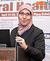
Biography:
She is an Assistant Professor of Endodontics at GMU, Ajman, UAE. She was a lecturer at the Department of Endodontics Faculty of Dentistry, Suez Canal University, Egypt. She got her Bachelor of Oral Surgery & Dental Medicine (BDS), Faculty of Dentistry, Alexandria University, Egypt in 1996. In 2003, she had her Master Degree in Conservative Dentistry (Endodontics) (MSc), Faculty of Dentistry, Suez Canal University, Egypt. Subsequently, she got her Doctoral Degree in Dental Sciences (Endodontics) (DDSc), Faculty of Dentistry, and Suez Canal University, Egypt in 2009. She is a member of many of the dental associations. She has several publications. She shared in various conferences as a speaker as well as a chairperson.
Abstract:
Bacteria have a major role in the success of an endodontic treatment. Mechanical preparation alone cannot effectively eliminate bacteria from dentinal tubules and other irregularities in the root canal. Few remnant micro‑organisms can multiply between appointments often reaching the same level as at the start of the previous session. These observations call for an effective intra‑canal medication that will help to disinfect the root canal system. Calcium hydroxide has an antibacterial effect on most of the endodontic pathogens. This is due to its high alkalinity. It damages the bacterial cytoplasmic membrane. Chlorhexidine has been found to have a broad-spectrum antimicrobial action and relative absence of toxicity. Calcium hydroxide points as well as chlorhexidine gutta percha points are ready to be used in a flexible way and easily to be introduced into the canal. Three bacterial cultures - before, after instrumentation and after medication by either one of the experimental materials - were taken from forty healthy patients having non-vital single rooted teeth with chronic apical periodontitis. Twelve types of bacteria were commonly isolated with different frequencies. Anaerobes represented about 56.1 % and aerobes represented about 43.9 % of the total bacteria. Streptococcus mitis (15.2 %) was the most frequent isolated bacteria (aerobes and anaerobes) from all the cases in the 1st culture. Then it was followed by Prevotella intermedia (14.7 %), Peptostreptococcus spp. (10.5%), Eubacterium lentum (9.4 %), Lactobacillus spp. (8.9%), Staphylococcus aureus (8.4 %), Veillonella parvula (7.9 %), Enterococcus faecalis (7.3 %), Escherichia coli (E-coli) (6.8 %), Neisseria catarrhalis (5.2%), Proionibacterium spp. (4.7 %), Staphylococcus epidermis (1 %). The results demonstrated a highly significant reduction in the bacterial growth in the experimental groups before and after instrumentation and medication of the root canal when compared to the control. Chlorhexidine gutta percha points proved to have a significant better antibacterial effect than calcium hydroxide gutta percha points.
Jules Poukens
Cranio-maxillofacial Surgeon Atrium/Orbis Medical Center, The Netherlands
Title: NEED A NEW SKULL OR MANDIBLE? 3D PRINT IT !
Time : 14:05-14:25

Biography:
Prof. Dr. Jules Poukens is currently lecturer and researcher at the Biomed Research Institute of the University Hasselt and the University of Leuven in Belgium. He is a Cranio-Maxillofacial Surgeon at the Medical Center Sittard /Heerlen in the Netherlands. He got his M.D. and D.M.D from the Catholic University Leuven in Belgium. He was trained as Cranio-Maxillofacial Surgeon in Belgium (Leuven) and Germany (Freiburg, Black Forrest). After his training, he first joined the staff at the University Hospital Maastricht and later on the staff of Medical Center Sittard /Heerlen in the Netherlands. He has participated in several European Community funded projects on medical rapid prototyping and manufacturing and was acting chairman of the Board of Directors of the EU funded project CUSTOM-IMD. He was the leading surgeon that designed and implanted the world’s first 3D printed total mandibular implant and was one of the pioneers in using 3D printed skull implants. He has numerous publications and held numerous presentations in this field. Born and raised in Belgium, Jules searched for his roots and lives with his wife and two daughters in Dilsen, Belgium near the Dutch and German border.
Abstract:
Patients in the cranio-maxillofacial clinic often present with serious, complex, and potentially life- threatening or life-limiting medical conditions (e.g. tumor, trauma, aggressive osteomyelitis). Available treatments may not always give satisfactory results for patients and doctors. Therefore, complex problems ask for new solutions. An emerging technique in the medical field is Computer Aided Design (CAD) , Computer Aided Manufacturing (CAM) by 3D printing. For successful implementation of CAD-CAM technology in the clinical practice doctors, dentists and engineers need to work together and share their expertise. This intense cooperation leads to 3D printing of custom patient specific implants. 3D printed implants are used for the treatment of skull defects, dental superstructures and world’s first 3D printed entire mandible replacement implant. These clinical cases will be highlighted.
Roula Al Bounni
Riyadh Colleges of Dentistry & Pharmacy, Kingdom of Saudi Arabia
Title: Direct composite veneers as a permanent treatment of anterior teeth
Time : 14:25-14:45
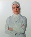
Biography:
Roula AL-Bounni currently is professor in operative dentistry. She has worked with undergraduate and postgraduate students in clinical & literature aspects in Operative & Endodontic department 1995-2011. She supervised more than 15 MSc and 6 PhD student’s certificates in Operative and aesthetic dentistry 2002 -2011. She was Head of Endodontic & Operative Dentistry Department 2007-2011
Abstract:
Every new material or technique introduced to the field of dentistry aims to achieve esthetic and successful dental treatments with minimal invasiveness. Therefore, direct laminate veneers have developed for advanced esthetic problems of anterior teeth. Tooth discolorations, rotated teeth, coronal fractures, congenital or acquired malformations, diastemas, discolored restorations, palatally positioned teeth, abrasions and erosions are the main esthetic problems for many patients, and it's basically the main indications for direct laminate veneers. Also, along passing time, Changes in color, shape, and structural abnormalities of anterior teeth might lead to important esthetic problems for patient, for that laminate direct composite veneers could be a good conservative solution for all these problems. Direct Laminate veneers are applied on prepared tooth surfaces with composite resin material directly in dental clinic. Absence of necessity for tooth preparation, low cost, intraoral polishing of direct laminate veneers is easy to use, and any cracks or fractures on the restorations may be repaired intra-orally, all of these are some advantages of this technique, however, the main disadvantages of direct veneers are low resistance to wear, discoloration and fractures, but the improvements of the new generations of resin composite restorations and adhesive techniques have surmount all these problems. But it's worth mentioning that the material itself, and the ideal application of this technique is the main factor to achieve long lifespan for successful outcome. By this technique we can get lot of benefits with wide range of people who seek improvements in there distorted smile due to numerous problems in anterior teeth.
Wisam Al-Rawi
University of Detroit Mercy School of Dentistry, USA
Title: The current state of Cone Beam CT (CBCT) 3D imaging in dentistry
Time : 14:45-15:05
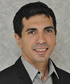
Biography:
Wisam Al-Rawi has completed his MSc degree in medical imaging (oral imaging option) from K.U.Leuven, Leuven, Belgium in 2006 and MS degree in oral and maxillofacial radiology from the University of North Carolina at Chapel Hill, NC, United States in 2009. He is currently holding an adjunct faculty position at the University of Detroit Mercy School of Dentistry and studying in the Accelerated Dental Program. He founded Marcilan Inc. A company dedicated for oral radiology education and created two educational apps in oral radiology for iPhone and iPad: CBCT and iPanoramic. He helped two dental schools to move to digital radiography. He has published several papers regarding Cone Beam CT and oral radiology education.
Abstract:
The introduction of Cone Beam CT (CBCT) constitutes a paradigm shift in the way clinicians acquire and view radiographs. Unlike intraoral or panoramic radiographs, there is no magnification and no superimposition of structures. CBCT volumetric imaging provides unprecedented highly detailed radiographic information about the patient’s teeth, bone, jaws, and airways spaces. This information can be used for diagnosis and treatment planning including implant planning, detection of root fractures, assessing the relationship of an impacted wisdom tooth to the mandibular canal, sleep apnea studies, growth assessment, pre and post-surgery assessment, and pathology. However, with CBCT, there is increased financial costs and radiation exposure to the patient. It is important that clinicians understand the advantages and limitation of this technology for better diagnosis and treatment planning of their patients. With many machines available on the market and more coming each year, it can clearly be seen that this technology is taking a big role in the field of dentistry. This purpose of this presentation is to: (i) review principles of action of CBCT; (ii) compare radiation dose of different machines; (iii) highlight clinical cases for use of CBCT in dentistry and how it can be used as a tool to aid in diagnosis and treatment planning of different cases in dentistry.
Carolina Duarte
Ras Al Khaimah College of Dental Science, UAE
Title: The potential of drug and gene therapy applied to orthodontic treatment
Time : 15:05-15:25

Biography:
Dr. Duarte did her pre-graduate training at the National Autonomous University of Honduras and obtained her PhD in Maxillofacial Orthognathics from Tokyo Medical and Dental University in Japan. She has studied the effect of hormones on bone metabolism and their application in orthognathic treatment, and presented and published her research internationally. She recently joined the faculty at Ras Al Khaimah College of Dental Science.
Abstract:
Interceptive and comprehensive orthodontic treatments are characterized by the use of appliances that mechanically modify bone size and shape, and reposition teeth in the dental arches. The bone modifications and tooth movements done with these orthodontic appliances can be complex and painful, require long retention time and are subject to significant amounts of relapse. Orthodontic research has focused on optimizing the mechanics, materials and esthetics of orthodontic appliances which have made orthodontic treatment more predictable and acceptable to patients. However, less importance is given to therapeutic adjuvants that may optimize bone remodeling during orthodontic treatment and improve hard tissue adaptation after treatment is completed. Numerous reports have been published on the effects of drugs or hormone therapy during orthodontic treatment, as well as the genes involved in bone remodeling during orthodontic tooth movement. This basic knowledge could be translated to safe clinical therapies with the potential to reduce treatment and retention time, facilitate anchorage and control relapse. Gene therapy is trending in many areas of medical and dental practice but has been overlooked by orthodontists. It is only logical to wonder if orthodontic treatment will continue to grow as a purely mechanic therapy or will evolve to a hybrid practice using mechanics and gene therapy to optimize its progression and results.
Lamis Abuhaloob
Ministry of Health, State of Palestine
Title: School dental health in Palestine
Time : 15:25-15:45

Biography:
Lamis abuhaloob obtained PhD in Dental Public Health from Newcastle University in the United Kingdom. She is Head of Oral and Dental School Health in Ministry of Health in Palestine. She has published and presented several papers in national and international journals and conferences of dentistry.
Abstract:
By the end of 2012, Palestinians (including refugees) living in Palestinian territories were 5.8 million, approximately, 40% of them were school children. Since the eve of the 1948 war, the Occupied Palestinian Territories have suffered from political conflict, resulted in serious deteriorations in the socio-economic status and psycho-social wellbeing of the Palestinian people. World Health Organization stressed the high burden of oral diseases and its adverse effect on general health and quality of life. Several studies found that oral diseases have profound impact on children’s performance in schools, nutrients intake, and growth. Despite, school dental health programme in Palestine have been established to promote general and oral health among school children in government and UNRWA schools, there was a gradual increase in the prevalence of dental disorders and mainly dental caries. Generally, the def was higher than DMF. Incidences of periodontitis, fluorosis and malocclusion were the highest in the year of Al-Quds Intifada upraise, and in 10th grade schoolchildren. Increased oral and dental diseases in children in Palestine have overloaded the health services and caused a negative impact on the effectiveness of prevention programs and health promotion strategies in both of Palestinian Ministry of Health and UNRWA. Political unrest had a profound negative effect on oral health in Palestine. The increase in rate of decayed teeth in schoolchildren is alarm and suggests an increase in risk of dental caries in future generations. School dental health programme struggles to improve child oral and dental health. Thus, It should be revised. Effective oral health promotion interventions should consider age specific oral diseases and political unrest.
Coffee Break 15:45-16:00 @ Al Diyafah Pre-function area
Meheriar Chopra
Dr Meheriar’s Dental Clinic, India
Title: Lasers in root canal sterilization: A clinical case presentation
Time : 16:00-16:20
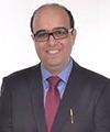
Biography:
Meheriar Chopra has completed his BDS and MDS in Conservative Dentistry and Endodontics from Maharashtra University of Health Sciences Ministry of Health, M.A.R Dental College, Pune, India. He has completed his Diploma in Lasers from SOLA (Society of Oral Laser Applications) affiliated to University of Vienna. He is a leading Endodontist with a private practice in Pune, India. He has research articles published in both national and international journals.
Abstract:
Lasers (Light amplification by stimulated emission of radiation) are slowly but surely revolutionizing dental treatment. In dentistry, lasers are accurate, preserve more healthy tooth structure, produce minimum to no pain during treatment, sterilize tissues with minimal to no bleeding and promote faster tissue healing. The first use of lasers in endodontics was in 1971. With a rapid development of laser technology, new lasers with a wide range of characteristics are now available. In endodontics they are used for a variety of procedures such as alleviating dentinal hypersensitivity, pulpal diagnosis, pulp capping and pulpotomy, cleaning and shaping of root canal systems, canal sterilization and endodontic surgery. The following clinical cases demonstrate the use of a diode laser in canal sterilization, promoting faster periapical healing. Studies have shown that lasers have a bactericidal effect. The laser beam as it diverges has a higher penetration depth enabling irradiation of the curvatures and complexities within the root canal system like lateral and accessory canals, fins, webs and apical deltas. Laser irradiation has the ability to remove debris and the smear layer from the root canal walls following biomechanical instrumentation. This enables the clinician to confidently clean and shape the root canal system in minimum time leading to higher patient satisfaction.
Vivek Padmanabhan
Ras Al Khaimah College of Dental Sciences Ras Al Khaimah, UAE
Title: Stress Estimation in Pediatric Dentistry – A Correlation Study between Modified Dental Anxiety Scale and Salivary Cortisol Levels
Time : 16:20-16:40

Biography:
Vivek Padmanabhan, B.D.S., M.D.S., (PhD), Assistant Professor, Department of Pediatric Dentistry, Ras Al Khaimah College of Dental Sciences, Ras Al Khaimah University of Health Sciences, Ras Al Khaimah, UAE; Tel: +971527400018; e-mail: drvivekpadmanabhan@gmail.com 1 Department of Pediatric Dentistry, Ras Al Khaimah College of Dental Sciences, Ras Al Khaimah University of Health Sciences, Ras Al Khaimah, UAE. 2 Department of Pediatric Dentistry, Bangalore Institute of Dental Sciences, Bangalore, India. 3 Department of Pediatric Dentistry, A.B.Shetty Memorial Institute of Dental Sciences, Mangalore, India
Abstract:
Anxiety is a frequent problem among dental patients in general and especially children. The presence of dental anxiety in pediatric patients is not a dilemma for the patients alone but also for the dental professionals themselves; and sometimes it renders the treatment more complicated and tedious to be accomplished successfully. Fear and anxiety increase the activity of the Hypothalamic Pituitary Axis (HPA) which in turn enhances secretion of cortisol. Cortisol also known as the stress hormone, is secreted from the adrenal cortex and dispersed to all body fluids, and can be detected in urine, serum or saliva. Heightened cortisol levels are thus indicative of increased stress as a result of elated fear and anxiety. Anxiety scales have been used for the assessment of anxiety in children. Most successfully used anxiety scale is the modified dental anxiety scale (MDAS).
120 children who required dental procedures were included in the study of which 60 each belonged to the study and control groups. For each of these children saliva was collected to evaluate salivary cortisol levels and MDAS was duly filled with the help of the parents to evaluate the anxiety levels.
The results indicated that the salivary cortisol levels and anxiety levels were significantly increased in the study group when compared to the control group. A positive correlation can also be seen between the two measurement methods of anxiety levels.
Keywords: Anxiety, Stress, Cortisol, MDAS
Jadranka Handzic
University hospital center, Croatia
Title: Tympanometric findings in cleft palate patients: influence of age and cleft type
Time : 16:40-17:00
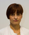
Biography:
Jadranka Handžić graduated in 1984y. at Medical School University of Zagreb, took Master degree at 1987y. And PhD at 1989. Since 1987y she spent her residential program of Otolaryngology than diploma at 1989 as specialist of Otolaryngology and 2003y as sub-specialist of Audiology. At 2000-2001 she spent academic year on Fulbright Scholarship at Cleft Palate-Craniofacial Centre and Dental School of Medicine, University of Pittsburgh and Children’s Hospital Pittsburgh U.S.A Department for Paediatric Otolaryngology, University of Pittsburgh, U.S.A at position as Adjunct Associate Professor of Oral Medicine and Pathology and at 2001-2002 as Visiting Assistant Professor at Department of Oral Medicine and Pathology, Cleft Palate-Craniofacial Centre, Dental School of Medicine, University of Pittsburgh, U.S.A. 2001-2002 she had Lester Hamburg- Research Fellowship in Department for Paediatric Otolaryngology Children's Hospital of Pittsburgh, Medical School University of Pittsburgh, U.S.A. From 2002 she was Assistant Professor of Otolaryngology, University Clinical Hospital Centre and Medical School “Zagreb†and from 2008 Professor of Otolaryngology and Audiology. She is author and co-author of 16 articles in Current Contents, lecturer of 30 presentations on International conferences, reviewer in Journal and Annals of Maxillofacial Surgery, Director of 7 post-graduate studies of Otolaryngology and Audiology, author and co-author of 4 books, author of 3 international projects.
Abstract:
Objective: Conductive hearing loss associated with otitis media with effusion is usually finding in cleft lip and palate children. Method: Tympanometry performed in 57 children with bilateral (BCLP), 122 unilateral cleft lip and palate (UCLP) and 60 children with isolated cleft palate (ICP) according to age sub-groups, median age of groups 5y. All children undergo no previous ear surgery and undergo standard method of cheilognathopalatoplasty. Results: B type is most frequent in ICP (58.3%) less in UCLP (50.6%) and lowest in BCLP (46.5%).UCLP children show higher decrease B type ears with aging (rs=-0.4430) than BCLP (rs=-0.3186) and ICP (rs=-0.3378). Moderate hearing ears (21-40dB) showed highest decrease of B type with aging, lower decrease (rs=0.2184) have ears with mid threshold 11-20dB and lowest showed ears with severe hearing loss. At age 1-3y UCLP have higher rate of B type ears than BCLP and ICP which start to decrease at age 7-12y.At BCLP B type ears increase in frequency at age 4-6y.In ICP patients decrease in B type is not significant until 15y when first decrease showed. Type A is typical for older ages while B type for younger ages with significant decrease at age 4-67 and 7-9y no ear side difference. BCLP showed increase at 4-6y and the slowest decrease than other types. Conclusion: Cleft types due to craniofacial characteristics with aging have different mode of pathophysiological changes of middle ear desease.
Abeer Abo El Naga
King Abdulaziz University, Saudi Arabia
Title: Dentinal hypersensitivity: recent trends in the treatment
Time : 17:00-17:20
Biography:
Associate Professor and course director of third year Operative Dentistry at Faculty of Dentistry, king Abdulaziz University. Received my B.Sc. of Oral and Dental Medicine (1988), MSc in Restorative Dentistry, and PhD in Operative Dentistry (2007) from Faculty of Oral and Dental Medicine, Cairo University. I Served as Assistant Professor of Operative Dentistry at Misr University for Science and Technology (Cairo, Egypt) and Ibn Sina National College for Medical Studies (Jeddah, Saudi Arabia) from 2007 to 2011. I had presented and published more than 20 papers in reputed journals.
Abstract:
Dentine hypersensitivity is one of the most encountered problems in the dental clinics. When a patient presents with dentine hypersensitivity symptoms, he/she should be examined and informed of the proper treatment option to eliminate the problem. However, many dentists have some problems in diagnosing and treating dentine hypersensitivity. Selection of the most appropriate treatment option to achieve successful management is based on proper diagnosis for the condition and its etiological factor. This presentation will cover the following: the etiological factors, diagnosis and invasive vs. most recent non invasive treatment of dentine hypersensitivity.
Mohammad Abdulwahab
Kuwait University, Kuwait
Title: The Need for Anesthesia and Sedation Services in Kuwaiti Dental Practice
Time : 17:20-17:40

Biography:
Mohammad abdulwahab has completed his dental anesthesiology and publich health training from University of Pittsburhg and postdoctoral studies in managment of special needs dental patients from the same institute. He served as a faculty if univeristy of pittsbugrh school of dental medicine. Currently he is a faculty member at kuwait university faculty of dentistry. He a member in number of comittes and a mentor for residents. His research foucus on medical emergency and pain control. He has published in reputed journals and has been serving as consultante for training.
Abstract:
The public health relevance of the prevalence of dental fear in Kuwait and the resultant barrier that it creates regarding access to dental care was assesded. An analysis demonstrated a high prevalence of dental fear and anxiety in the Kuwaiti population and a perceived need for anesthesia services by dental care providers. The assessment of the general population showed nearly 35% of respondents reported being somewhat nervous, very nervous, or terrified about going to the dentist. In addition, about 36% of the population postponed their dental treatment because of fear. Respondents showed a preference to receive sedation and anesthesia services as a means of anxiety relief, and they were willing to go to the dentist more often when such services were available. People with high fear and anxiety preferred to receive some type of medication to relieve their anxiety. In conclusion, the significance and importance of the need for anesthesia services to enhance the public health of dental patients in Kuwait has been demonstrated, and improvements are needed in anesthesia and sedation training of Kuwaiti dental care providers.
Mustafa DAG
Gulhane Military Medicine Academy, Turkey
Title: The Maxillary Sinus Membrane Elevation Technique Using Venous Blood Without Grafting Material And Immediate Implant Placements: A Case Series Analysis
Time : 17:40-18:00
Biography:
Mustafa DAÄž graduated from Ankara University Faculty of Dentistry in 2007. He began his doctoral studies at Oral and Maxillofacial Department of Gulhane Military Medicine Academy in 2011 and is still ongoing. He is working especially on implant placement and bone augmentation topics. He has published more than 5 papers in national or international journals. He participated in many national and international congresses with oral or poster presentations.
Abstract:
Introduction: Various sinus augmentation techniques have been successfully used in order to enable the implant placements in the atrophic posterior maxilla. The object of the study was to evaluate whether sinus membrane elevation technique using venous blood without any grafting material constitute an effective technique for the maxillary sinus augmentation. Method: Eighteen patients who have not sufficient bone for implant placement at maxillary posterior sides were included in the study. Twenty-eight dental implants were inserted to twenty-two maxillary sinus just after the sinus membrane elevations via lateral approaches. After implant placements the cavity between relocated schneiderian membrane and sinus inferior wall was filled with venous blood without any bone grafting material. Cone beam computerized tomography was used for follow-up evaluation. Results: Comparisons of pre- and postoperative radiographs clearly demonstrated new bone formation between the new and former sinus inferior walls. Within the mean follow-up period of 1.5 years two implant failures were recorded. One of the failures was occurred just three months after the surgery due to infection, and the other implant failure was recorded almost a year after the prosthetic rehabilitation. The survival rate was nearly % 93 in the study. In a systematic review by Wallace et al, it was reported that average survival rate of implants placed in maxillary sinuses augmented with the lateral window technique was nearly %92. It was shown that the technique used in this study has provided high implant survival rate as studies using grafting materials for maxillary sinus augmentation. Conclusion: The case series of 18 patient demonstrated that the maxillary sinus membrane elevation procedure without grafting material and immediate implant placements is considered to be more cost-effective and less time-consuming compared with conventional procedure.
Sultan Aldeyab
Ministry of National Gauard, King Abdulaziz Medical City, Riyadh
Title: Correction of Excessive Spaces in the Esthetic Zone
Time : 18:00-18:20
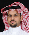
Biography:
Sultan Aldeyab has completed his AEGD certificate with honor in 2008, and then he got Saudi Board of Restorative Dentistry with honor from 2008-2012. He is working in Restorative Department in King Abduaziz Medical City (National Guard) Riyadh, He is teaching restorative post graduate resident and lecturing undergraduate student in dental collage of King Saud Bin Abdulaziz University for Health Sciences. He got many awards and he has many publications and lectures.
Abstract:
Introduction:
The use of porcelain crowns and veneers to solve esthetic problems has been shown to be a valid management option especially in the anterior esthetic zone. This case report discusses a patient having diastema in the anterior region. The patient was treated with orthodontic treatment and porcelain crowns & veneers in the maxillary arch for the closure of diastema.
Clinical report:
A 35 year old male patient with a chief complaint of discolored anterior teeth and gaps between the teeth. The patient was unhappy with the appearance of his teeth.After thorough examination; impressions for diagnostic models were made in irreversible hydrocolloid. The models were studied to decide the shape and size of the restorations with help of a diagnostic wax up. Before proceeding for tooth preparation, shade was selected using Classical shade guide (Vita Zahnfabrik, Germany). The maxillary teeth were then prepared from right 2nd premolar to the left 2nd premolar to receive porcelain crowns and laminate veneers. Impression of the maxillary arch was made in addition silicone.
The laminates were etched with 4 % Hydrofluoric acid, after etching, they were washed thoroughly using liberal amount of water. On drying, a coat of Silane coupling agent (Porcelain Primer, Bisco,USA ) performed on two teeth at a time starting at the midline.
The prepared teeth were etched using 37% Phosphoric Acid for 15 seconds. On air drying bonding agent was applied & light cured for 10 seconds. Composite luting agent was used for cementation. The laminates were spot cured for 5 seconds initially. Excess cement was removed with explorer and then complete curing was done for 20 seconds. On completion of the cementation procedure, the occlusion was checked in centric and eccentric positions for interferences. The high points were removed and polished.
Discussion:
The etiology of diastema may be attributed to the following factors: (a) Hereditary- congenitally missing teeth, tooth and jaw size discrepancy, supernumerary teeth & frenum attachments; (b) Developmental problems- habits, periodontal disease, tooth loss, posterior bite collapse (Oesterle & Shellhart, 1999). Treatment planning for diastema correction includes orthodontic closure, restorative therapy, surgical correction or multidisciplinary approach depending upon the cause of diastema (Dlugokinski et al, 2002).
Conclusion:
Orthodontic treatment, bonded porcelain crowns & veneers can provide successful esthetic and functional long-term service for patients.
- Track 15: Oral Implantology
Track 16: Prosthodontics
Track 18: Dental Surgery
Location: Al Dhiyafah 5-6

Chair
Hasan Alkumru
University of Western Ontario, Canada

Co-Chair
Ioannis Georgakopoulos
University of Modena & Reggio Emilia, Italy
Session Introduction
Ioannis P. Georgakopoulos
University of Modena & Reggio Emilia, Italy
Title: IPG-DentistEdu Technique Minimal Invasive Sinus Bone Augmentation without Sinus Floor Elevation with Intentional Perforation of the Sinus Membrane using the Autologous Biomaterial CGF-CD34+Matrix
Time : 09:30 - 09:50

Biography:
Prof. Dr. Ionians P. Georgakopoulos BPP University, School of Health, London-UK BPP University, School of Health, London-UK. Faculty member. Professor at BPP University, Clinical Master in Implantology & Oral Surgery (Director Prof. Maher Almasri) University of Modena & Reggio Emilia, Italy. PhD School “Enzo Ferrari†Faculty of Mechanical Engineering & Biomaterials. Dental Materials & Dental Prosthetic technologies. Assistant Professor.
Abstract:
Aim: The rapid placement of implants in the sinus cavity by means of intentional perforation of the sinus membrane following a certain clinical protocol, without performing Sinus Floor Elevation (SFE). Materials & Methods: 32 patients with age range between 43-67 (19-female, 13-male), in which upper jaw rehabilitation needed to be performed with nonremovable prosthesis. The option of placing a total of 47 implants (28 left and 19 right on sinuses sides) was offered to the patients. All of them have been informed regarding the clinical procedure and a written consent was signed. This study has undergone an ethics review from Patras University. According to the proposed clinical protocol, all implants were placed in a flapless approach and entered each sinus cavities with intentional perforation of the Schneiderian membrane. The combined employment of concentrated growth factors (CGF & stem-cells-CD34+) and bone grafting within the osteotomy site and by means of implant immersion, was made in such a manner that the sinus can adapt to the new conditions forming new bone around the implants without the need to perform an SFA procedure. Results: CBCT scans showed new bone formation around the implants by means of textural image analysis. None of all patients’ sinuses presented any signs of infection. Implant Stability Quotient values ranged between 61 and 69 proving high implant strength. Histologic analysis showed alternate layers between non-Mineralized Tissue and Vital Bone. Conclusions: IPG-DentistEdu technique promising results demonstrate that it can be considered as a reliable alternative to the SFE (Sinus Floor Elevation) procedure.
Ren-Yeong Huang
National Defense Medical Center, Taipei, Taiwan
Title: Maxillary Sinus Floor Elevation via Crestal Approach: the Hydraulic Pressure Technique
Time : 09:50-10:10
Biography:
Ren-Yeong Huang has completed his DDS, periodontology training and PhD degree in oral biology and immunology from National Defense Medical Center. He is currently an assistant professor in periodontology and the director of the advanced implant center at the Tri-Service General Hospital. He is also a diplomate of the Taiwan Academy Board of Periodontology and Implantology, and serves as editorial board member and ad hoc reviewer for peer-review journals. Dr. Huang’s strong interest in clinical- and basic- oriented topics, including pathogenesis of periodontal disease, bone regeneration and dental implant therapies has led to his extensive publication in peer-reviewed journals.
Abstract:
For many clinicians, inadequate alveolar bone height and anatomical features of the maxillary sinus complicate sinus lift procedures and placement of endosseous implants. We review the recent advance in sinus elevation and present a series of cases using the new internal crestal approach that addresses these issues and. A new device for maxillary sinus membrane elevation by the crestal approach using a special drilling system and hydraulic pressure, and placing dental implants placed after a sinus lift procedure. Our experience suggests that hydraulic sinus condensing is a predictable and minimally invasive alternative for prosthetic rehabilitation of maxillary anterior and posterior regions in the presence of anatomical restrictions to implant placement.
Manish Agrawal
Bharti Vidhypeeth University , India
Title: Occlusal appliance for TMD
Time : 10:25-10:45
Biography:
Dr. Manish Agrawal is a Consulting Prosthodontist & Implantologist. He did his Graduation and Post-graduation from Mumbai University India. He is a skilled Prosthodontist, very well known for his clinical skills. He is a Professor and Postgraduate teacher at Bharti Vidhypeeth University Pune India. He is a Director of Smile Dental Studio and Zenith Dental Laboratory. He has presented papers at National & International levels, and has various publication to his credit .He excels in interdisciplinary dentistry, with Special emphasis on Full mouth rehabilitation & smile designing. He has being awarded the best Prosthodontist of the year 2014.
Abstract:
The term Temporomandibular Joint (TMJ) Diseases and Disorders refers to a complex and poorly understood set of conditions. They usually manifest as pain in the area of the jaws and their associated muscles, frequently accompanied by limitations in the ability to make normal jaw movements. Treatment of occlusion related TMJ disorders is often a challenge for both the dentist and the patient. These disorders can be difficult to diagnose as the presenting symptoms can be variable. Conventional occlusal splint therapy is a safe and effective, albeit conservative mode of treatment in comparison to the surgical therapies for temporomandibular joint disorders (TMD). Once the cause of the occlusion related disorder is identified, this reversible, non-invasive therapy not only yields diagnostic information but also provides relief to the patient, without the difficulties that often accompany other, more complex approaches. The goal of this presentation is to familiarize “physicians with the masticatory system†and to delineate the basic principles of occlusal splint therapy for treating temporomandibular disorder (TMD), bruxism and some forms of headaches.
Rivan Sidaly
Oslo University School of Dentistry, Norway
Title: Hypoxia increases the expression of enamel proteins and cytokines in an ameloblast-derived cell line
Time : 10:45-11:05

Biography:
Rivan Sidaly has completed her Master’s degree in Dentistry at the age of 24 years from Oslo University and continued on to earn her doctoral degree at the same university. She is researching on the relationship between hypoxia and molar incisor hypomineralization.
Abstract:
The aim of the study was to investigate the effect of hypoxic conditions on the expression of enamel proteins, and secretion of alkaline phosphatase (ALP), cytokines and interleukins from an ameloblast-derived cell line. The murine ameloblast-derived cells (LS-8) were exposed to 1% oxygen concentration (24 or 48 h) and harvested after 1, 2, 3 and 7 days. The effect of the hypoxic condition on the gene expression was measured by qRT-PCR. The secretion of cytokines and interleukins, and the alkaline phosphatase (ALP) activity into the cell medium was measured by Luminex and a colorimetric assay, respectively. The effect was calculated relative to the expression and secretion from untreated cells (controls) at each timepoint. Hypoxic exposure for 24 and 48 hours increased the expression of the structural enamel matrix proteins; amelogenin (Amel), ameloblastin (Ambn), enamelin (Enam), and the enamel protease matrix metallopeptidase-20 (MMP-20). Hypoxia-inducible factor 1-alpha (Hif-1a) mRNA expression increased also after both 24 and 48 h exposure to hypoxia. Similarly several vascularization factors (monocyte chemotactic protein 1 (MCP-1), and vascular endothelial growth factor (VEGF)) and pro-inflammatory factors (interleukin-1 alpha (IL-1α), interleukin-1 beta (IL-1β), interleukin-6 (IL-6), interleukin-10 (IL-10), interferon-gamma (INF- γ), and Interferon gamma-induced protein 10 (IP-10)), were also increased by 24 and 48 hours of hypoxia. ALP activity was down regulated by both 24 and 48 hours of hypoxia, whereas the LDH level in cell culture medium was higher after exposure to 24 hours compared to 48 hours of hypoxic conditions. These results indicate that hypoxic exposure may disrupt the controlled fine- tuned expression and processing of enamel proteins, and promote the secretion of pro-inflammatory factors.
Feras Aalam
Al-Noor Specialist Hospital, MOH, Saudi Arabia
Title: Timing of implant placement after extraction: Immediate, Early or Delayed
Time : 11:05-11:25
Biography:
Feras Aalam Board Member, Saudi Fellowship in Dental Implant Scientific Committee. Holder of Hajj Medal from the Minister of interior for the Excellence in providing a positive impact on the success of the Hajj season.Honor degree for the Excellence in training during residency on the Saudi Board in Advanced Restorative Dentistry program and Consultant in Implantology and Restorative Dentistry Program Director of Saudi Board of Advanced restorative Dentistry Makkah Dental Center, Al-Noor Specialist Hospital and his publications are Characterization of heat emission of light-curing units
Abstract:
Restorative therapy performed on implant(s) placed in a fully healed and non-compromised alveolar ridge, has high clinical success and survival rates. Currently, however, implants are also being placed in sites with ridge defects of various dimensions, fresh extraction sockets, the area of the maxillary sinus, etc. Although some of these clinical procedures were first described many years ago, their application has only recently become common. Accordingly one issue of primary interest in current clinical and animal research in implant dentistry includes the study of tissue alterations that occur following tooth loss and the proper timing thereafter for implant placement. In the optimal case, the clinician will have time to plan for the restorative therapy (including the use of implants) prior to the extraction of one or several teeth. In this planning, a decision must be made whether the implant(s) should be placed immediately after the tooth extraction(s) or if a certain number of weeks (or months) of healing of the soft and hard tissues of the alveolar ridge should be allowed prior to implant installation. The decision regarding the timing for implant placement, in relation to tooth extraction, must be based on a proper understanding of the structural changes that occur in the alveolar process following the loss of the tooth (teeth).
Shady A M Negm
Pharos University, Egypt
Title: Common Implant Mishaps and Failures due to practitioners careless procedures or lack of knowledge regarding implantology
Time : 11:25-11:45
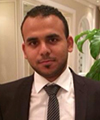
Biography:
Shady Negm graduated dental faculty. He then completed a one year of training in Implant Dentistry at Egyptian Society of Oral Implantologists. He joined a large group practice in Alexandria, Egypt from 2010- 2014 during which he served as a Board Member and a Core Dentist for the American Journal of Biomedicine. He is a fellow of the Alexandria Oral Implantology Association (AOIA). He has been awarded Diplomat status in the Implant Dentistry from Seville University, Spain. He serves as the Continuing Education Implant Course Director at the Dental Town Magazine, United States. He is an editor in International Journal of Dental Clinics (E-ISSN 0975-8437, P-ISSN 2231 – 2285) since October 2014 till now. Currently, Dr. Negm serves as a Full-Time Faculty Member at the Faculty of Dentistry, Pharos University.
Abstract:
Depending on our long time practice in the field of implant, We would give you some tips regarding implant failures and mistakes which might happen. The reason of developing so many dental implant mistakes is because of implant dentistry was not a part of the dental school curriculum of the vast majority of dentists in practice today. Only now are dental schools starting to incorporate adequate training in implantology. So that, there are many reasons that mistakes happen. Many dentists simply don’t possess the training or qualifications necessary to be successful. Sadly, these dentists are more concerned with saving money by cutting corners or performing a procedure too quickly. Below we have provided several factors that can contribute to dental implant mistakes. And we will illustrate the Common Reasons for Dental Implant Failure; Many dentists use two-dimensional panoramic x-rays to place dental implants. Although this method works well for most dental surgeries, there is much more sophisticated technology available for dental implants. So we have to use 3-D CT scans which give a much clearer Figure of the exact position of nerves and blood vessels present in the bone. These powerful CT scans combined with radiography techniques are used to best determine the precise placement of every dental implant. They only take a few minutes and radiation exposure is minimum. Another main reason for dental implant failure is the quality of the fixture. With over two hundred companies that provide dental implants, there are only a handful of reputable companies with proven research that documents their reliability and quality. The Temptation is great for dentists to save on costs with cheaper fixtures. Costs vary greatly with substandard products, coming in nearly one-one hundredth of the cost of high quality fixtures. We are learning now that cutting on costs can lead to serious complications and problems for the patient. Substandard materials can be used to save costs. Also many complications can result like infection, nerve damage that causes facial numbness and pain, or the implant can be misplaced into the sinus cavity.
Gaurav Singh
AMU, India
Title: Immediate Loading Revisited With the Concept of Intra-Oral Welding
Time : 11:45-12:05
Biography:
Dr. Gaurav Singh has completed his BDS from Karanataka University, India in 1996 and MDS in Proshtodontics & Oral Implantology from Rajiv Gandhi University of Health Sciences, Bangalore in 2001. After completing his Master’s degree he is working in the department of Prosthodontics, Dr. Z.A. Dental College, AMU., Aligarh till date. He is working as Associate Professor in the department. He has many articles, published in both National and International Journals.
Abstract:
In implant dentistry, it has been claimed that process of Osseointegration requires on an average undisturbed healing of three months in mandible and six months in maxilla. To decrease this time the concept of immediate loading was introduced, initially many clinicians were doubtful about immediate loading. They advised that immediate load exerted at implant interface may interfere with the process of bone healing, lead to implant failure. But, many clinical and in Vitro trials have shown that long term success of removable and fixed prosthesis of immediate loaded implants can be achieved. Bone quality and quantity may play significant role. Beside accurate presurgical diagnostic and treatment planning, implant Macro & Micro design, the adequate fixation and immobility of the Implant are of utmost importance to prevent the risk of micro movement related to surrounding bones. A high predictability of immediate implant loading with fixed provisional restoration has been shown in many reports.
All previously describe technique for reinforcement of acrylic resin provisional restorations involve either the use of thin wire or fibers throughout the span, or a time consuming fabrication of a cast metal framework. The main objective of this presentation is to introduce a prosthetic concept for an accelerated rigid splinting of multiple implants for same day immediate loading with metal reinforced acrylic resin provisional restorations by utilizing the Syncrystallization technique.
Mayyadah Al-Mozainy
King Saud University, Saudi Arabia
Title: Characterization of Polymerization Shrinkage and Water Absorption of Low-shrinkage Light-polymerized Composite Resin Materials
Time : 12:05-12:25
Biography:
Mayyadah Al-Mozainy, Professor from Saud University, Saudi Arabia, Doctor Mayyadah Al-Mozainy is a speaker at Dentistry-2015
Abstract:
Aim of this study: to compare the polymerization shrinkage and the water sorption values of different low shrinkage composite materials. Materials and method: different commercially available composite resin materials were used in this study: Reflexions XP (BISCO, IL, USA), Kalore (GC CORPORATION, JP -Tokyo), AELITE LS (BISCO, IL, USA), Filtek P90 (3M, St. Paul, USA) and Tetric N-ceram (Ivoclar Vivadent, NY, USA). The data for water absorption were analyzed by two-way analysis of variance (ANOVA), followed by one way analysis of variance (ANOVA) and Tamhane test at 95% level of confidence. Results: Tetric N-ceram showed the highest polymerization strain mean ( -12.52 ± 10.958 ) followed by Filtek P90 (-10.99 ± 9.188), Reflexions XP (-8.77 ± 6.662 ), GC Kalore ( -5.20 ± 3.185 ), and the least polymerization strain was recorded for Aelite LS ( -3.95 ± 2.717). Filtek P90 showed the highest mean growth (5.21 ± 5.71) among the tested materials. Comparing the mean of water absorption percentages of the tested materials to each other, Filtek P90, Aelite LS and GC Kalore showed no significant difference to each other with P=0.986. Aelite LS , GC Kalore and Reflexions XP were also not significant to each other with P=1.00. Tetric N-ceram and Reflexions XP were not significantly different from each other with P=0.411, although Tetric N-ceram was significantly different from all the other tested materials with P=0.000. Conclusion: The results of the present study showed that the polymerization shrinkage for all the tested materials was the greatest during the light activation reaction and decreased after the curing light was turned off, and they all presented their highest readings during the first twenty seconds. The highest polymerization shrinkage strain was recorded for Tetric N-ceram followed by Filtek P90, Reflexions XP, GC Kalore and Aelite LS.
T.S. Vinoth kumar
Sri Ramachandra University, India
Title: Influence of cryogenic treatment on hardness of Nickel Titanium alloy used for rotary endodontic instrument – An Invitro study
Time : 12:25-12:45
Biography:
T.S. Vinoth kumar has completed his MDS at the age of 25 years from The Tamilnadu Dr MGR Medical University. He is the research scholar in the Department of Conservative Dentistry and Endodontics, Faculty of Dental Sciences, Sri Ramachandra University. He has published national and international papers in reputed journals and has been serving as invited reviewer and editorial board member. He coined the term â€Reverse Sandwich Restoration†in dentistry.
Abstract:
Cryogenic treatment is a one time supplementary thermal treatment where alloys are subjected to sub-zero temperature to improve their metallurgical properties. Nickel Titanium alloy used for manufacturing of rotary endodontic instruments possess superelastic property due to which they must be machined rather than twisted. The combination of surface defects during manufacturing and lower hardness of the alloy results in compromised cutting efficiency of endodontic instruments within the root canals. A recent approach deals with the use of Nickel Titanium alloy that possess a mixture of austenite and martensite phase at body temperature in order to have extreme felxibility without the conventional shape memory. The pupose of this study was to evaluate the effect of different cryogenic treatment parameters on the hardness of Nickel Titanium alloy. Accordingly, a similar virgin alloy was procured in the form of sheets. 15 samples were prepared from the sheet using electrical discharge maching process. They were randomly divided into five groups (four experimental groups based on the soaking time and temperature parameters of cryogenic treatment and a control group; n=3). The metal samples in each experimental group were subjected to vickers hardness testing following the cryogenic treatment and compared with that of the control group. The data were tabulated and the results were statistically analysed using SPSS software.
Aitor de Gea Rico
Whipps Cross Hospital, London
Title: Infective endocarditis of presumed dental origin and the NICE guidelines: An updated overview
Time : 12:45-13:05
Biography:
Aitor de Gea Rico graduated from the University of Barcelona, School of Dentistry in 2005 where he received award of merit in 12 subjects including Oral Surgical Pathology. He received his equivalent to Masters of Dental Science from the same university in 2007. Aitor moved to the United Kingdom to work as a General Dental Practitioner within the National Health Service for 3 years. While working as a Senior House Officer in Oral & Maxillofacial Surgery (OMFS) in Bristol and Birmingham (UK), he obtained the Diploma of Membership of the Dental Faculty of the Royal College of Surgeons of Edinburgh in 2010. He was awarded the Ivor Whitehead Prize, by the British Dental Association West Midlands Hospitals Group in May 2011 for an oral presentation. Aitor graduated in Medicine from Barts and the London School of Medicine, University of London, in 2014. He also worked as a Clinical Fellow in OMFS at Kings College Hospital and Queens Hospital in London while undertaking his studies. He is currently in his first year of Foundation Training at Whipps Cross Hospital, London, as part of his continued training to pursue a career in OMFS. Aitor has achieved a number of publications in peer-reviewed journals and has presented some of his work at a national and international level. He is a member of the British Medical Association and British Association of Oral and Maxillofacial Surgery and has developed a particular interest in the treatment of patients under intravenous sedation.
Abstract:
Introduction: Antimicrobial prophylaxis against Infective Endocarditis (IE) in patients undergoing interventional procedures is no longer recommended in the UK. The National Institute for Health and Care Excellence (NICE) Guidelines published in 2008 has been object of significant controversy within the scientific community. A number of recently published case reports add on argument to the debate. We present a case of IE following dental treatment for which NICE guidelines were followed.
Case report: A 59-year-old gentleman presented with a 1 week history of general malaise, nausea, loss of appetite and lethargy after minimal exertion. Known mitral valve prolapse was reported, and an initial differential diagnosis included subacute bacterial endocarditis. An echocardiogram and Streptococcus mitis growth in the blood cultures supported the likely diagnosis. He was referred to the Oral and Maxillofacial Surgery Department where his previous dental history revealed dental treatment 1 month before admission. In line with the current guidelines, the patient did not receive antibiotics pre-operatively.
Discussion: Dental professionals should be able to recognise the signs and symptoms of IE. It is important to remain informed about such a pathological entity and be aware of its diagnosis and management. Patients with certain cardiac conditions who are at increased risk of developing IE still remain at risk, despite the recent change in the NICE guidelines. The promotion of preventive dentistry and medicine is crucial to reduce the overall risk of IE. The incidence of IE since this radical change in practice remains to be quantified and act upon. Involving patients in informed decision-making and individualization of cases when antibiotic prophylaxis is considered seem to be a more ethical approach.
Walaa KM Hafez
Cairo University, KSA
Title: Evaluation of marginal bone stability around immediate implants
Time : 13:45-14:05
Biography:
Walaa KM Hafez from Cairo University, KSA, made his valuable remarks at International Conference & Exhibition on Dentistry
Abstract:
Aim: The aim of the study was to evaluate marginal bone changes following immediate implantation in the maxillary anterior region.
Patients & Methods: This study was conducted on 20 adult patients indicated for extraction & immediate implant insertion in one or more of the upper anterior teeth. The peri-implant defect was filled with mixture of autogenous bone (collected from the chin) & PRF (platelets rich fibrin) and covered with PRF membrane to cover the implant site. The changes in the marginal bone were assessed at 0, 3, and 6 months using CBCT. All data were collected and statistically analyzed. The data were presented as means and standard deviation (SD) values. Wilcoxon signed-rank test was used for the changes in mean defect height, width and area after 6 months. Spearman’s correlation coefficient was used to find out the correlation between clinical and radiographic bone height in cross-section.
Results: The marginal bone was stable in 83% of cases with 6 month follow up period. Through the whole study period (Immediate – 6 months); the radiographic height showed statistically non-significant decrease at the mesial side. Distally and cross-sectionly, there was a statistically significant decrease in mean defect height. As regards the defect depth, the change in depth was limited to the 2 mm bone margin.
Conclusions: Particulate autogenous bone - PRF mix is effective in obliterating the buccal bone defect around immediate implants in the maxillary anterior region. The marginal bone was stable in 83% of cases with 6 month follow up period.
Marco Roy
Poznan University of Medical Sciences, Poznan Poland
Title: Photofunctionalization: A New Method to Bio-Activate the Titanium Implant Surface
Time : 14:05-14:25

Biography:
Marco Roy has completed his dental degree at the Poznan University at the age of 24 years. He is attending a postdoctoral study at Poznan University of medical sciences in the field of dental implantology. He is an Assistant Professor in the department of Implant prosthodontics. He has published in reputed journals and has been actively participating in research groups.
Abstract:
Titanium implant surfaces inevitably undergo biological aging, associated with lower bone-implant-contact (BIC) values. Already 4 weeks from manufacturing the implant surfaces are contaminated with polycarbonyls and hydrocarbons that modify their physio-chemical state, leading to significant reductions of BIC values (45-75%). and, likely, low numbers of stem cells actually reaching the surface of the fixture. However, photofunctionalization (UV light irradiation) of the surface can reverse the biological aging of titanium, recovering BIC values virtually to 100% and greatly enhancing the osteointegration process. In our study we investigated how UV irradiation may affect osteogenic environment, increasing cell adhesion, migration, proliferation and differentiation. First, we evaluated how the changes in surface molecular composition affect hydrophilicity and electrostatic state of titanium and how it is correlate with UV intensity. Second we tested the hypothesis that the bio-activated titanium surface may attract more stem/progenitor cells and/or promote their osteogenic differentiation. To this aim, human osteogenic cells seeded onto bio-activated or untreated surfaces were compared at different time points for cellular and molecular properties. Particular attention was focused on the expression profiles of regulatory gene networks (HOX and TALE) known to reflect positional, embryological and hierarchical identity of human stromal cell populations, thus providing useful biological markers.
Abhinav Gupta
AMU, India
Title: Guidelines and rationale behind Abutment selection and designing in Implant Prosthodontics
Time : 14:25-14:45

Biography:
Dr. Abhinav Gupta has completed his BDS and MDS in Prosthodontics and Oral Implantology from Manipal Academy Of Higher Education, Manipal, India . He is a consultant and senior Assistant Professor in Department of Prosthodontics, Aligarh Muslim University, Aligarh. He has worked in a govt. funded research project in the field of oral implants at Maulana Azad institute of Dental Sciences, New Delhi. He has completed one year advanced training in oral implantology from DGOI Germany. He has research articles published in both national and international journals.
Abstract:
The implant evolution has become a "restorative driven" field and it is therefore important to know the designing principles of abutments and its connection to the implants and its rationale for use in clinical practice. During the past several decades, there has been a significant increase in the number of dental implant manufacturers and implant restorative components available for clinicians and dental laboratory technicians. Selecting the appropriate abutment can be both complex and confusing with the ever-increasing number of implant choices and transepithelial abutments available. Many restorative dentists resort to fabricating costly custom abutments to avoid the selection process. Although custom abutments are at times necessary, prefabricated abutments are usually more desirable. This article will describe the various abutments available and how to select the correct abutment for a given clinical situation in an organized, systematic fashion. Criteria discussed include implant position, angulation, soft tissue height, and interocclusal space. The latest modifications and developments in implant abutments are reviewed along with an indirect method of selecting abutments in a laboratory setting.
Nicola Luigi Bragazzic
University of Genoa, Italy
Title: Ramadan fasting and oral pathologies
Time : 14:45-15:05
Biography:
He was born on the 2nd of March in 1986 in Carrara (MS), Tuscany (Italy) and is currently a MD, a MSc, a PhD and a resident in Public Health. He got his MD (medical degree) on the 15th of July in 2011 with a final mark of 110/110 cum laude with a thesis on Personalized Nanomedicine ("Nanomolecular aspects of medicine, at the cutting-edge of the nanobiosciences in the field of health-care") and the joint Italo-Russian MSc (Master of Science) in nanobiotechnologies at Lomonosov Moscow State University (27th April 2012). He got his PhD in nanochemistry and nanobiotechnology at Marburg University, Germany with a final mark of “very good†and is currently a resident in Public Health at University of Genoa, Italy, 3rd year. He is author and/or co-author of several publications.
Abstract:
Ramadan fasting represents one of the five pillars of the Islam creed. Even though patients are exempted from observing this religious duty, they could be willing to take part into the religious ceremonies. Here, we review the extant literature focusing on the impact of Ramadan fasting on patients suffering from oral pathologies. From the collected evidences, we can claim that: 1) trans-cultural counseling of patients suffering from oral diseases is extremely important; 2) Muslim subjects could experience malodour and halitosis; the exact etiology of this phenomenon is complex, due to the accumulation of sulphur-containing compounds in the oral cavity, a decrease in salivation and changes in the oral microflora. An accurate oral hygiene when breaking the fast is recommended, for example using miswak which has anti-bacterial properties; 3) dental operations can be performed using special precautions, changing drugs and administering intramuscular or trans-dermal treatment instead of oral agents; appointments can be delayed or postponed, if necessary and possible; 4) patients with chronic systemic diseases, and especially metabolic disorders, such as diabetes, should take care of their oral cavity; 5) mouthwash and mouth-rinsing without water swallowing are allowed practices in Islam and ameliorate athletic performances, even though some patients or subjects could be reluctant to do it, perceiving these practices as a break of the fast.
- Advanced Course

Chair
Fabio Savastano
Preseident, ICNOG, Italy
Session Introduction
Fabio Savastano
International College of Neuromuscular Orthodontics and Gnathology, Italy
Title: Neuromuscular orthodontics: The future is here
Biography:
Fabio Savastano is a Medical Doctor from Italy with over 25 years of experience in Orthodontics and Gnathology. He did Postgraduate Master in Orthodontics from the University of Padua, Italy, founded Neuromuscularorthodontics.com (now ICNOG) in 1994. He has lectured in India, Middle East, Canada, Brazil and Italy and he is President of the International College of Neuromuscular Orthodontics and Gnathology (ICNOG). He practices in Albenga, Italy, and is limited to orthodontics and TMD.
Abstract:
The key in understanding occlusion and malocclusion is the analysis of function. Neuromuscular Orthodontics is a relatively new and easy approach engineered for the orthodontist of the future. This brief course has the objective to introduce you to the basics of Neuromuscular Orthodontics: Muscle function (EMG), lip balance, cranio-cervical posture, mandibular tracking, muscle relaxation (T.E.N.S.). During this course you will be shown several cases treated with a variety of functional appliances for the treatment of Class I-II-III malocclusions and oral orthotics for the treatment of TMJ disorders. See how easy treatment is once your comprehension of the cause of malocclusion is exhaustive. Part 1 of the course will focus on how to diagnose a malocclusion without all the complicated tasks that have been taught in the past, but in a more modern and functional fashion. Part 2 will show you how to build a better looking face while reducing relapse and TMJ problems.
Why should they attend?
1. This course will open your mind as an orthodontist. Understanding the hidden functional characteristics of the individual is a must in planning an ideal treatment.
2. Where is the mandible supposed to be relative to the cranium? The correct cranio-mandibular relationship guarantees an optimal TMJ function: A key to success.
3. See mandibular advancement in growing and adult patients: Build beautiful faces.
- Track 1: Basic Dentistry
Track 2: Dental Biomaterial Science
Track 4: Diagnosis and Prevention of Oral Disease
Track 7: Orthodontics and Oral Health
Location: Al Dhiyafah 5-6

Chair
Anas H. Al-Mulla
European University College, UAE

Co-Chair
Kamis Gaballah
Ajman University, UAE
Session Introduction
Anka Letic
Rakcods, Ras Al Khaimah, UAE
Title: Use of Drugs and Biomaterials Important to Significantly Improve Overall Quality of Dental Work
Time : 12:25-12:45

Biography:
Prof. Letic-Gavrilovic Anka, DDS, PhD, duringlong scientific career collaborated with Royal Postgraduate Medical School, Histochemistry Department, London; Fukuoka Dental College, Department of Biochemistry, Fukuoka, Japan. Now works as Assoc.Prof. of Physiology and Biochemistry at RAKCODS, Dental College, United Arabic Emirates. Researching inoral biochemistry, immunology and basic clinical mechanism of dental implantology, Dr Anka expressed particular scientific interest in Biomaterials for bone reconstruction. Most of the work was dedicated to the regulatory role of growth factors in morphogenesis and postnatal development of the immune system and salivary glands. Recently, Dr Ankais very active in field of brain neurophysiology and practical applications. Neuromarketing is an academic approach to better understand how information leads to changes in attention, emotional responses, preference formation, choices and learning. Dr Letic’s present interest remains beautiful mystery of brain function. While there’s general agreement that attention and emotional engagement of brain, can be tracked, identifying specific emotions with confidence has been elusive. Therefore, data from neuromarketing are still best used when triangulated with more traditional data such as interviews, questionnaires, and historical data.
Abstract:
A smile is a very powerful social instrument. A better quality of working life, together with the promotion of employment and entrepreneurship, requests beauty and time savings. Today, patients come to Dental office and expect quick, good quality, long-term smile and absolutely, and always with no pain or other complications during or after the treatment. The successful Dentist is the one to be able to prepare and finish dental procedure; having happy patient at the and! Pharmaceutics could help a lot, but specific knowledge is needed.
This course is not meant to be an exhaustive compendium of pharmacology. New generations of healthcare professionals must be ready for inter-professional collaboration, as well as to be prepared to treat growing needs of patients for new type of treatments. Technological advances in health care industry every day deliver new pharmaceutical products that dentist can use and serve patients. Today, most presenters of aesthetic dentistry only show their exceptionally good work on teeth and gums while neglecting the complex face and/or lips. Without treating the face, the aesthetic work is incomplete unless the patient on their own sees other doctors. Without treating the face, your good work in the mouth will appear incongruous. Therefore, this course focuses primarily on those drugs and biomaterials used by the Dentist that highly improve results of dental work, boost doctor’s endeavor and significantly upgrade reputation of the dental office. These drugs are: 1) Pain killers, 2) Antialergics and antioxidants, 3) Skin and Lips fillers and 4) Bone Grafting Biomaterial. All these drugs and synthetic biomaterials will be listed, reviewed in terms of their clinical indications and applications, particularly choosing clinical requests, explaining mode of work, their clinical advantages and disadvantages comparing to other options, in terms of appropriate usage, adverse side effects and potential interactions.
Learning Objectives:
1. Understand the actions of and appropriate therapeutic use of pain killers, oral sedatives, local anesthetics, and controlling anxiety by medications;
2. Understand the rational use of anti-alergic agents in dentistry, both in terms of the management of existing orofacial allergies and for prophylaxis against the new developments;
3. Understand the importance of bone function, defective status in altering the absorption, distribution, metabolism, and therapeutic action of drugs such as Bisphosphonates’ or natural and synthetic bone graft substitutes;
4. Understand the bottom line whether Botox and dermal fillers are within the scope of dental practice for use by general dentists for dental esthetic and esthetic dental therapeutics.
A.M. Elamin
College of Sustainability Sciences and Humanities, UAE
Title: Putative periodontopathic bacteria and herpes viruses interactions in the subgingival plaque and whole saliva of patients with aggressive periodontitis, healthy controls
Time : 12:45-13:05
Biography:
A.M. Elamin is well known for his research publications.His keen research is on pathology of dental sciences.Presently he is working as professor at College of Sustainability Sciences and Humanities, UAE.
Abstract:
Background and Objectives: The microbial profile of aggressive periodontitis patients is considered to be complex with variations among populations in different geographical areas. The aim of this study was to assess the presences of four putative periodontopathic bacteria (Aggregatibacter actinomycetemcomitans, Porphyromonas gingivalis, Tannerella forsythia, Treponema denticola) and two periodontal herpes viruses [Epstein-Barr virus-1 (EBV), human cytomegalovirus (HMCV)] in subgingival plaque and whole saliva of Sudanese subjects with aggressive periodontitis and healthy controls. Material and Methods: The study group consisted of 34 subjects, 17 aggressive periodontitis patients and 17 periodontally healthy controls (14-19 years of age). Whole stimulated saliva and pooled subgingival plaque were collected and analyzed for detection of bacteria and viruses using loop mediated isothermal amplification (LAMP). Results: Prevalence of subgingival A. actinomycetemcomitans, HCMV and P. gingivalis were significantly higher among aggressive periodontitis patients than periodontally healthy controls. Co-infection with A. actinomycetemcomitans, HCMV and/or EBV-1 was restricted to the cases. Pooled subgingival plaque showed significant higher prevalence of A. actinomycetemcomitans and P. gingivalis than whole stimulated saliva in this population (p=0.0001, p=0.04). Increased risk of aggressive peritonitis was the highest when A. actinomycetemcomitans & was detected together with EBV-1(OD 49.0, 95% CI 2.5-948.7, p= 0.01) and HCMV (OD 39.1, 95% CI 2.0 - 754.6, p= 0.02). Conclusions: A. actinomycetemcomitans and HCMV were the most associated test pathogens with aggressive periodontitis in this population. Saliva may be useful as an alternative sampling tool for selected pathogens. However, parallel subgingival sampling is recommended for A. actinomycetemcomitans and P. gingivalis.
Lunch Break 13:05-13:45 @ Al Tanour Restaurant
Ala Al-Dameh
Specialist Endodontist Private practice, UAE
Title: Current advances in irrigation
Time : 13:45-14:05
Biography:
Ala Al-dameh BDS, Doctor of Clinical Dentistry (DClinDent), graduated in Dentistry from the University of Otago in New Zealand, after which she workedin private and government sectors in both New Zealand and overseas.She then served on the Faculty of Dentistry in the Department of Oral Rehabilitation at the University of Otago, and obtained her Doctorate in Clinical Dentistry in Endodontics. Her research interests include both clinical and laboratory research with a focus on the microbiology and disinfection of root canal dentine. As a side line she invests time on new instrumentation devices in root canal therapy. She is now Assistant Professor of Endodontics at Dubai College of Dental Medicine.
Abstract:
The role of bacteria in the development of pulp and periapical disease has long been established. Successful root canal therapy depends on thorough debridement of pulpal tissue, dentine debris and microorganisms. Irrigation has a key role in successful endodontic treatment. It kills planktonic and biofilm bacteria, dissolves and removes tissue, and helps files cut dentin in a safer and more effective manner.The aim of this presentation is to develop a clear understanding of the role of irrigation, particularly apical irrigation, and the effect of various solutions on dentine and biofilm. A safe and effective irrigation protocol for root canal treatment will be presented. Learning Objectives: • Develop a clear understanding of the role of irrigation particularly apical irrigation in successful endodontic treatment • Understand the effect of various irrigants on dentine and biofilm • Understand the interactions between the various chemicals found in irrigants • Develop a rational irrigation protocol for root canal treatment
Erika Lander
Private Educational Center Somos Saludy Educacion, Venezuela
Title: From simple to complex in oral rehabilitation
Time : 14:05-14:25
Biography:
Department of aesthetic Dentistry,Private Educational Center Somos Saludy Educacion,Caracas, Venezuela;Universadad Nacional Experimental politecncas de la Fuerza armand of caracas, Venezuela;Department of operative and Esthetic Dentistry,Universidad Santa Maria of caracas, Venezuela.
Abstract:
It is not always simple if it seems simpleâ€. In the absence, loss or tooth fractures, it gets open in our time an “endless†possibility of treatments using different options and restorative materials. We can evaluate “the simpleâ€, and “the complexâ€, i.e. from simple to complex in oral rehabilitation not based on the material used, but in the restorative value of each individual case. Which is important is a successful end.It is a resume of the different treatments (going from the simpler to the more complex alternative) to restore and rehabilitate the patients. From veneers and endocrowns to full mouth implants.
Chiquit van Linden van den Heuvell
University Medical Center Groningen, Netherlands
Title: Dental gagging problems? Train and relax!
Time : 14:25-14:45
Biography:
Chiquit van Linden van den Heuvell works as a clinical psychologist at the University Medical Center Groningen, initially at the Department of Medical Psychology, beginning in 2003 at the Department of Oral and Maxillofacial Surgery and Special Dental Care. She is member of the Regional Disciplinary Board for Health Care Psychologists in Groningen. Her psychotherapeutic activities concern patients with dental anxiety, dental gagging and medically unexplained symptoms. Research activities are mainly directed at Quality of Life in Head and Neck Oncology, with special focus on qualitative research into ethical and existential aspects of decision making in this field. In order to stimulate a more meaningfully discussion with patients about the question “Will it be worth it?â€, she started interviewing treated patients and next of kin of deceased patients, asking them: “Has it been worth it?â€. Another field of research interest concerns the development of a measurement instrument for dental gagging.
Abstract:
Dental gagging can be a burden in many respects. Implant retained dentures may be considered the solution for edentulous patients suffering from dental gagging. But the process of applying these devices may reveal gagging problems to such an extent that special approaches will be necessary. For dentate patients dental gagging may impede proper oral care, oral treatment or the wearing of a partial prosthesis. A ‘treatment of choice’ seems to be lacking, since research on dental gagging is almost exclusively restricted to case studies. More basically, reliable and valid procedures for evaluating any treatment do not exist. So notwithstanding the prevalence and inconveniences of dental gagging, knowledge about etiology, incidence and treatment is minimal. At the Center of Special Dental Care of the University Medical Center Groningen, the Netherlands, dental gagging is a field of interest since more than 10 years. A diagnostic instrument has been developed and a weekly multidisciplinary office hour for dental gagging patients has added considerably to our expertise, and it still does. This has resulted in a multidisciplinary method that will be described and illustrated with video fragments. Pros and cons of implants with dental gagging will be clarified. Recommendations and specific tips will also be provided for specialists who want to transcend the level of home remedies for their gagging patients. The core issues of this presentation are firstly that gagging patients seem to benefit from learning to become ‘an expert’ in the awareness of an approaching gag reflex, secondly, that dental implants are not a solution for EVERY patient, thirdly that removable dentures are preferable to dental implants if possible, and finally that the implantology trajectory should be preceded or accompanied by a training to enhance the patient’s control over the gag reflex.

Biography:
Hellen has worked as dental hygienist, in both the public and private sector and as a clinical tutor. She volunteers internationally on regular work trips to Vietnam and held industry leadership and advocacy roles, including Presidency of peak body, the Dental Hygienists’ Association of Australia Inc. (DHAA) and a member of the National Minimal Intervention Dentistry Workshop and Alliance for Caries Free Future. Hellen is currently completing a Master degree in Clinical Leadership and residing in Dubai
Abstract:
In the development of professional citizenship, clinicians may appreciate the community is an extension of our own clinical practice. We embrace our patients for whom we provide excellent clinical care in our everyday practices, patients with appointments and waiting for treatment and also extend our professional obligations to those who one day may seek dental treatment and anyone now or in the future who can benefit from treatment and community health promotion.
Practitioners developing professional citizenship may have the opportunity as individuals to be involved in community health promotion or clinicians may join professional societies or seek leadership roles and experience participating in collaborative partnerships and coalitions. Societies may provide professional development education, mentoring, and foster research and publish guidelines for evidence-based practice for the profession. Leaders may be decision- makers tackling the challenging aspects of health and normative economics. (What should we do with resources and what do our societal values tell us is fair and equitable)
Efficiency and equity is as integral to Australia’s heath care system as it is to the health care systems of the Arab Nations. However has become increasingly challenging to achieve the equitable distribution of limited resources within a mixed health care system and impact on Australia’s health inequalities among the homeless, rural and regional residents and indigenous Australians. Targeted vertical equity dental schemes present challenges when delivered via the private sector present have their own set of challenges including addressing equitable access in rural and remote areas, dynamic efficiency of the health care system and allocative efficiencies.
Kamis Gaballah
Ajman University, UAE
Title: Minimizing the Injury of Inferior Dental Nerve during Removal of Lower Third Molar: A Systematic Approach
Time : 15:05-15:25
Biography:
Dr. Kamis Gaballah is currently Associate Professor in Oral and Maxillofacial Surgery at Ajman University, UAE. Since graduation on 1993 Dr. Gaballah showed a great interest in the field of Oral and Maxillofacial Surgery for which he was extensively trained and practiced in many countries including Libya, Egypt, Ireland, United Kingdom, Norway and UAE. During his surgical training he was awarded with the fellowship of the Royal College of Surgeons in both England and Ireland . Dr. Gaballah was also formally trained in Oral Medicine at the Trinity College Dublin of the Ireland. Up on his completion of the professional surgical training and practicing for several years in the field, he has joined the University of London for intensive and novel research on the cancer gene therapy. His research work focused on the use of the tissue engineering, virus-mediated gene therapy for head and neck cancer and oral potentially malignant lesions and also the inhibition of angiogenesis to prevent the recurrence of head and neck cancer in surgically treated patients. He has published his novel research outcome in highly-ranked journals and his papers were cited by significant number of authors all over the world. His academic interest was expanded to include the higher education teaching and learning for which he was formally trained at the University of London and warded with membership of the UK council of higher education before joining the King’s College London as a Clinical Lecturer in Oral Surgery.
Abstract:
Worldwide, the presence of impacted third molar is often associated with significant morbidities that may range from soreness , pain, swelling, inability to open mouth widely & chew properly & impairment of the health of adjacent teeth to more serious complication like neck infection & emergence of certain pathologies like cysts & tumors. This wide range of clinical problem made the surgical removal of these teeth are the commonest oral surgical procedure. This procedure is associated with significant morbidity including pain & swelling, together with the possibility of temporary or permanent nerve damage, resulting in altered sensation of lip or tongue. This talk will shed some light on the systematic approach may be considered to prevent or minimize the damage to the inferior dental nerve during such surgeries.
Abeer AlSubait
Ministry of National Guard, Saudi Arabia
Title: Assessment of radiographic and oral manifestations in adult Saudi patients with renal failure and its relation to duration of renal failure and dialysis
Time : 15:25-15:45

Biography:
Dr. Abeer AlSubait got her Master Degree in Public Health, King Saud Bin Abdulaziz University for Health Science (College of Public Health and Health Informatics) 2012. She got her Diploma degree in Public Health from Liverpool Tropical Medicine 2012, AEGD certificate from King Abdulaziz Medical City-Dental Center – Jan 2004. She is the Director of School Dental Prevention Program in Ministry of National Guard, Riyadh, Saudi Arabia. She is Co-Director of the Advanced General Dentistry Program in Dental Service in Ministry of national Guard, Riyadh, Saudi Arabia. She is the Co-chairman of continuing education committee in dental service in Ministry of national guard , Riyadh, Saudi Arabia and Lecturer in Dental College in King Saud Bin Abdulaziz University for health science.
Abstract:
The number of patients with renal failure disease on hemodyalisis (HD) is increased and they require special attention and care from dentists, usually patients with end stage renal disease have another medical conditions as Diabetic, Hypertension, immunosuppression, using antihypertensive or anti coagulant or antiplatelet medications, making the dental treatment more complex , these patients develop a lot of oral and dental problems such as enamel hypoplasia, enamel attrition, periodontal disease, drug induced gingivitis, oral infections, ulcerations, and bone alterations due to hyperparathyroidisim, osteoporosis, narrowing of pulp chamber, osteosclorosis of the roots and abnormal bone healing after extraction, therefore dentists must be aware about these manifestations that may occur due to renal diseases. Dental care among these patients and preventive measures seems to have been neglected, and dentists are not aware about the oral, dental and bone manifestations that may occur as a result of renal failure, therefore the aim of this study is to measure the prevalence of radiographicbone and oral soft tissues and teeth manifestations among Saudi adult patients with renal failure on HD, and to find the relationship between the duration of renal failure/dialysis and the appearance of bone, soft tissue and teeth manifestations.
Andre Wilson Machado
Federal University of Bahia, Brazil
Title: 10 commandments of smile esthetics
Time : 15:45-16:05

Biography:
Andre Wilson Machado, D.D.S., M.S., PhD is an Associate Professor for an orthodontic program, School of Dentistry, Federal University of Bahia, Brazil. He is also a Visiting Professor at University of California Los Angeles (since 2011). He completed his dental education at School of Dentistry, Federal University of Bahia, Brazil and orthodontic education at Pontifical Catholic University of Minas Gerais, Brazil. He completed his Master’s degree at the Pontifical Catholic University and his PhD at the Paulista State University – Araraquara, Brazil. His thesis research work has been published in various international journals, not necessarily limited to orthodontics. He also is an author and co-author of more than 60 articles published worldwide. He has given lectures in more than 30 cities, totaling over 100 presentations. His current focus has been establishing protocols for smile esthetics diagnosis and treatment. Recently, he developed the smile analysis protocol entitled “10 commandments of smile esthetics†and also the “digital smile planningâ€, a brand new method to diagnosis and planning esthetic treatments in simple software such as Powerpoint and Keynote. He is a member of the Brazilian Association of Orthodontics and of the World Federation of Orthodontics and the actual scientific director of the Brazilian Association of Orthodontics – Section of Bahia.
Abstract:
The demand for esthetic treatments has become routine in health professional offices and for the public worldwide. Consequently, the primary reason patients seek dental treatment is to improve their smile esthetics. Therefore, the aim of this presentation is to introduce the smile analysis protocol entitled: “10 commandments of smile estheticsâ€.
Munad J. Al-Duliamy
AL_Iraqia University, Iraq
Title: Enhancement of orthodontic anchorage and retention by the local injection of strontium: An experimental study in rats
Time : 16:05-16:25
Biography:
Department of Orthodontics, College of Dentistry / AL-Iraqia University, Baghdad. Munad J. Al-Duliamy, Assistant Lecturer, Department of Orthodontics, College of Dentistry / AL-Iraqia University, Baghdad, IRAQ.
Abstract:
Objectives: To examine the clinical and histological effects of locally injected strontium on the anchoring unit of a rat model of an experimental relapsed tooth movement. Materials and methods: Thirty-six 10-week-old male Wister rats were randomly divided into two groups of 18 animals that were then randomly divided into three subgroups of six animals corresponding to three observation periods: T1 = 1 week, T2= 2 weeks, and T3 =3 weeks. In the first experiment, both the right and left maxillary first molars were moved buccally with a standardized expansive spring. Strontium chloride solution was injected every 2 days into the subperiosteal area buccal to the left maxillary first molar (the experimental side). The right-sided first molar was injected with distilled water as a control. In the second experiment, maxillary first molars were moved buccally with the spring. After 3 weeks, the spring was removed. Two days before the spring removal, strontium chloride was injected into the palatal side of left-sided maxillary first molar and distilled water was injected into the palatal side of the right-sided maxillary first molar as in experiment1. Results: At the end of the experimental period, significant levels of inhibition were noted in terms of both tooth movement and relapse movement in strontium-injected sides. Histological examinations showed that strontium enhanced the number of osteoblasts and reduced the number of osteoclasts.
Coffee Break 16:15-16:30 @Al Diyafah Pre-function area
- Symposium and Workshop (11:25-12:25 & 16:40-18:40)
Location: Al Dhiyafah 5-6
Session Introduction
Mutlu Özcan
University of Zurich ,Switzerland
Title: International Symposia on Direct adhesive bridges and splints using fibers: Step by step procedures

Biography:
She has authored more than 250 scientific and clinical articles in peer-reviewed journals, has given over 400 presentations at international scientific meetings, is a frequent lecturer at scientific meetings, receiver of several international awards and has held numerous continuing education courses in Europe. She serves also for the editorial boards of several scientific journals. Her clinical expertise is on reconstructive dentistry. She has also Visiting Professor Positions at various universities including São Paolo State University (Brazil), Federal University of Juiz de Fora (Brazil), University of Brno (Czech Republic), University of Madrid (Spain) and University of Florida (USA).
Abstract:
Durable adhesion of glassy matrix or oxide-based ceramics is crucial especially for minimally invasive reconstructions. This lecture will highlight the fundamental principles of adhesion to different ceramics, cover current knowledge and the clinical protocols regarding to surface conditioning methods and adhesion promoters to be used in conjunction with different resin-based materials.
Learning objectives:
1- Prerequisites for durable adhesion to different ceramics
2- Surface conditioning methods and working mechanisms
3- Clinical sequence of adhesion protocols for cementation and repair
German Ramirez-Yanez
Private Practice, Ontario, Canada
Title: Myofunctional orthodontics: The future for preventing, intercepting and treating malocclusions
Biography:
Ramirez graduated in 1986, obtaining his D.D.S. degree from the Javeriana University in Colombia. Afterwards, he completed a Pediatric Dentistry Diploma in Mexico. Dr. Ramirez interest includes guiding craniofacial growth and development in children. Thus, he trained in treating bite problems in Brazil and completed a Doctoral degree (PhD) in Oral Biology in Australia. Beside his extensive experience treating children, Dr. Ramirez has been an academic in USA and Canada. He became a fellow of the Royal College of Dentist of Canada in 2012 and recently moved to Ontario, where he practice, while continue associated with the Faculty of Dentistry, University of Manitoba in Winnipeg, Canada. He is also a fellow of the Royal College of Dental Surgeons of Ontario and a member of the Ontario Dental Association and the American Academy of Pediatric Dentistry. He is a member of the editorial board of the Journal of Dentistry and Oral Health and the Journal of Orthodontic Science and Practice. He is the author of the book “Early Treatment of Malocclusions: Prevention and Interception in Primary Dentitionâ€, as well as co-author of the book “The Trainer System: A myofunctional approach to treat malocclusionsâ€. He investigates on Craniofacial Growth and Development, the Patho-Physiology of Functional Disorders in the Cranio-Cervico- Mandibular system and how the craniofacial structures are modified by functional appliances.
Abstract:
Course description: The reason orthodontics is delivered mostly in permanent dentition and largely a cosmetic procedure is that we have been focusing on malocclusion as the problem to solve, much like we focus on any facial deformity that needs “correctingâ€. However, a shift in focus that looks at malocclusion as a symptom of imbalances elsewhere in the face, starting at an early age, will bring another perspective to the general and pediatric dentists to foreseeing malocclusions. By looking at the sources of imbalance we get a better picture of the etiology of malocclusion and a framework for treating it. In that way, malocclusions could be prevented and intercepted at an early age. We will explore the past, present, and possible future of early interventions as seen from this different point of view. This course will provoke you to think differently about diagnosis and treatment planning, especially as the goals of dentistry and medicine begin to merge. Course goals: To present insights on craniofacial growth and development and put myofunctional thinking in diagnosis and treatment planning on the cutting edge of early intervention of malocclusions. Course objectives: To Produce a Shift in Perspective: Just as Heisenberg taught us to look at light in two different ways, this lecture will illustrate a second, distinct way for the general and pediatric dentists to look at malocclusion. To Understand the Etiology of Malocclusion: The etiology of malocclusion is largely epigenetic. This lecture discusses what we are missing in our current concepts of “Growth and Development†and how a new understanding lets us vision malocclusion in a completely different way. To Envision the Mouth as Part of the Body: As our concept of malocclusion widens, so do our areas of concern and our scope of practice. This lecture will discuss some of the “big picture†health concerns related to the smaller issue of malocclusion. We will talk about what is behind the etiology of malocclusion, how a healthy airway is of major concern, and how our discipline interconnects with many other healthcare specialties. To Understand and Apply Myofunctional Orthodontics: Diagnosis depends on our ability to see what’s there. In this lecture we will learn how to “read†the body, the face, the skull, and the teeth in order to see the imbalances behind the malocclusion. Once the diagnosis becomes clear, the goals of treatment make sense. MFO treatment focuses on reversing the damage caused by poor function, improving oral-muscular function, and avoiding further damage, real prevention and interception.
- Special Session for Students (14:45-18:30)
Location: Al Dhiyafah 4

Chair
Anka Letic
RAK College of Dental Sciences, UAE
Session Introduction
Mehrdad Ghaffari Targhi
Dental University of Shahid Sadoughi-Fazaye sabz Blv, Yazd, Iran
Title: Story telling; a preferred strategy in oral health education
Time : 14:45-14:55

Biography:
Mehrdad Ghaffari Targhi currently is a dentistry student in Yazad University, Iran of Medical Science. He has been researching academically since 2011 and has strong research knowledge in the respective field.
Abstract:
Objectives:
Selection of appropriate educational strategy in the field of oral health is considered as an important issue in prevention of oral and dental diseases and as oral health promotion in community. Importance of this made us to compare two methods of storytelling ( indirect method) and lecturing(direct method) in oral health education of elementary school students in yazd.
Methods:
This intervention study was conducted on 117 female elementary school students in 1391-92. Random and clustered sampling was done from 4 groups of grade 3 in elematary school. data selection was through a questionare, so that knowledge, attitude and practice of students were evaluated and compared before and after administration of methods. Data were analyzed using SPSS- 17 software through T-test and ANOVA.
Results:
The mean knowledge scores were 15.59+ 1.94 and 16.96+ 0.79 in instructed group by lecturing and storytelling respectively and the difference between two groups were statistically significant ( p-value = 0.001). The mean attitude scores were 21.40+ 3.5 and 24.32+ 2.55 in instructed group by lecturing and storytelling respectively and the difference between two groups were statistically significant ( p-value = 0.001). The mean practice scores were 13.42+ 2.01 and 14.39+1 in instructed group by lecturing and storytelling respectively and the difference between two groups were statistically significant ( p-value = 0.003).
Conclusion:
Findings of this study indicated that storytelling method (indirect method) had more significant effect than lecturing method (direct method) on knowledge, attitude and practice in oral health education.
Keywords: health education, Storytelling, oral and dental hygiene
Nita Kumari Bhateja
Aga Khan University Hospital, Pakistan
Title: Overjet as a predictor of vertical facial morphology in orthodontic patients with class II division 1 malocclusion
Time : 1455-15:05
Biography:
Nita Kumari has completed her BDS in 2009 from Liaquat University of Medical and Health Sciences, Jamshoro Pakistan. Currently, she is doing FCPS-II training (fellowship of College of Physician and Surgeon Pakistan) in speciality Orthodontics at Aga Khan University Hospital and is in her final year of residency program.
Abstract:
This research aims to evaluate the vertical facial morphology in untreated orthodontic patients with Class II division 1 malocclusion. The sample comprised of 113 patients (61 females and 52 males) between 8 and 13 years of age, having Class II malocclusion with overjet of >4 mm, no prior history of orthodontic treatment, no craniofacial anomalies and no missing first permanent molars. Skeletal parameters were assessed by using pretreatment lateral cephalograms of these patients. Overjet was measured on the study casts taken from each subject using digital vernier caliper. Descriptive statistics were calculated for age and different vertical facial cephalometric angles. Pearson’s correlation was used to correlate various parameters. One-way ANOVA was used for comparison of means of vertical facial cephalometric angles among three overjet groups (Group-I = 5-7 mm, Group-II = 8-10 mm, Group-III = >10 mm). The means of all the vertical facial cephalometric parameters were in the normal range representing average facial pattern in patients with Class II division 1 malocclusion, except Jaraback ratio which indicated tendency towards long facial pattern. No statistically significant correlation was found between overjet and the parameters of vertical facial morphology. Frankfort mandibular plane angle was found to have moderately significant positive correlation with Steiner’s mandibular plane angle (0.789**) and Y-axis (0.604**). Patients with Class II division 1 malocclusion have an average vertical growth pattern. Overjet value is not a predictor of vertical facial morphology. There is no significant correlation between overjet and parameters used to assess vertical facial morphology
Monisha Singhal
Chandra Dental College, India
Title: Modern techniques in management of white spots of tooth enamel
Time : 15:05-15:15
Biography:
Monisha Singhal has done her BDS from Maharana Pratap Dental college and now pursuing her Posgraduation in Pediatric Dentirsty at Chandra Dental College, Lucknow. She is member of Indian Societry of Pedodontics & preventive Dentistry.
Abstract:
White spot lesions are frequently seen because of incipient caries, Developmental Defect of Enamel or post-orthodontic decalcification in the dental clinics. These lesions present esthetic problems as well as the progression of demineralization.. Various conventional treatments available i.e topical agents in form of creams, pastes and remineralizing agents such as fluoride therapy, casein phosphopeptide amorphous calcium phosphate pastes, bleaching therapy ,Microabrasion, conventional bonding and veneering agents.. DMG Icon is a new minimally invasive technique which works through resin infiltration on enamel defects. With DMG Icon, white spot lesions are stabilized while the anatomical shape and color of the noncavitated white spot defects of enamel are masked.
Syeda Mahvish Hussain
Aga Khan University, Pakistan
Title: Amount of tooth substance gained with crown lengthening: A Systematic Review
Time : 15:15-15:25
Biography:
Syeda Mahvash Hussain Assistant Professor, Operative Dentistry Aga Khan University.Validity of Different Dental Age Estimation Methods in Pakistani Orthodontic Patients.
Abstract:
BACKGROUND:
Extensive caries, short clinical crown, traumatic injury, or severe para-functional habits may limit the amount of tooth structure available to properly restore an affected tooth. The restoration of an adequate biological width and the creation of an adequate space for proper placement of crowns prosthetic margins on a compromised tooth can be achieved surgically (crown lengthening procedure) or orthodontically (forced eruption), or by a combination of both.
OBJECTIVE:
The purpose of this systematic review was to find out which crown lengthening procedure is the most commonly used and provides best results in terms of amount of tooth substance gained.
METHODS:
Search engines like Pub med and CINAHL plus (Ebsco) were used to search articles related to our review question using the key terms and different permutations:
• Surgical crown lengthening,
• Gingivectomy/gingivoplasty
• Biologic width,
• Orthodontic extrusion,
• Sub-gingival restoration
• Ferrule
RESULTS:
• The total number of teeth assessed in all 8 studies was 321 (range 20-84 per study)
• The surgical site was mentioned in 2/8 studies which made a total of 73 from the 321 teeth assessed.
• Only 80 of the 321 teeth assessed in the 8 studies were affected because of subgingival caries and another 53 had a fracture going sub-gingivally and the remaining studies did not mention the clinical presentation
• The radiographic evaluation was assessed in only 1/8 studies which had 30 teeth
• In 7 out of the 8 studies, (total of 291 teeth), did not mention which jaw maxillary or mandibular, they belonged to.
• The most common indication for crown lengthening in these 8 studies was for proper restorative treatment
CONCLUSIONS:
• The number of clinical trials on CLS (crown lengthening surgery) was limited.
• The quality of the studies which report data on CLS was mostly inadequate because basic demographics like the surgical site, the type of jaw (mandibular/ maxillary), radiographic evaluation and clinical presentation were missing from most of the studies evaluated
• APF (apical re-positioning of flap) with bone reduction was the most commonly used technique (7/8 studies) for CLS
• The mean amount of tooth structure gained initially was 2.46mm which decreased to 1.49mm after 6months.
Aisha Khoja
Aga Khan University Hospital Karachi, Pakistan
Title: Validity of Different Dental Age Estimation Methods in Pakistani Orthodontic Patients
Time : 15:25-15:35
Biography:
Dr. Aisha Khoja has completed her BDS in 2009 from Liaquat University of Medical and Health Sciences, Jamshoro Pakistan. Currently, she is doing FCPS-II training (fellowship of college of physician and surgeon Pakistan) in speciality Orthodontics at Aga Khan University Hospital and is in her final year of residency program.
Abstract:
This research aims to evaluate the validity of Demirjian’s (1973), Nolla’s (1960) and Willems (2001) methods of dental age estimation in Pakistani orthodontic patients (8-16.9years). It also addresses the validity of these methods in determining dental maturity across the gender and compares the difference between original Demirjian tables (based on French-Canadian standards) and tables formulated for Pakistani population by Sukhia et al. (2012). Orthopantomograms of 403 subjects (males: 176, females: 227) were examined for dental age assessment by different methods. Paired t-test and Wilcoxon signed-ranked test were used to determine significant differences between mean dental age (DA) and chronological age (CA) among different age groups. Correlations between DA and CA were assessed by the Spearman’s correlation (p=≤0.05). Nolla’s method under-estimated the DA in males (1.00±1.54) and over-estimated in females (0.21±1.64). DA was significantly advanced using Pakistani tables (males=0.32±1.17, females=0.38±1.33years) and Willems method (males= 0.31±1.09, females=0.29±0.48years). However, DA was better correlated with CA using Pakistani tables as compared to the French-Canadian standards. Earlier dental maturation was reported in girls than boys using Demirjian and Nolla’s methods. Strong and significant correlations were found between CA and DA according to all methods (p<0.001). Among all, Willem’s method was identified as the most valid method for dental age estimation in Pakistani orthodontic patients)
Rabia Ali
Aga Khan University Hospital, Karachi, Pakistan
Title: Comparison of Topical Analgesic and Saline Rinses in Post Extraction Healing among hypertensives and non-hypertensives
Time : 15:35-15:45
Biography:
Rabia Ali graduated from Aga Khan University Hospital, Karachi, Pakistan. Her research interests are in Dentistry, Event management and Marketing. She has given the opportunity to prepare the article on event published in the 2015 on “Comparison of Topical Analgesic and Saline Rinses in Post Extraction Healing among hypertensives and non-hypertensivesâ€
Abstract:
There is no evidence based guidelines on using saline rinses for post extraction oral care among hypertensives. Similarly, benefit of orally dissolved topical analgesics in addition to orally administered analgesic is questionable. This study was conducted to compare post dental extraction healing among subjects who took simple analgesic tablets dissolved in water versus those who used saline rinses post operatively. A study was done among patients who underwent dental extractions at AKU dental clinic, Karachi. Carious, periodontally mobile, traumatized or broken down teeth among 20-70 years old in either gender were included. In addition to routine prescription of antibiotics and analgesics, hypertensive subjects(n=20) were advised dissolved Aspirin tablet as an oral care while non-hypertensive were divided into two sub groups(n=20 each), advised saline mouth rinses or dissolved Aspirin for 5 days respectively. The outcome (healing of extraction socket) was evaluated 7 days post-extraction on ordinal scale. The mean age of our sample was 40.9±13.5 years. There were 50 females and 10 males. Upper left second molar was the most commonly extracted tooth(n=5). Out of 60 subjects, 5 were diabetics, all belonging to hypertensive group. Non-hypertensive subjects on topical aspirin showed the best socket healing(20/20) followed by hypertensives on Aspirin(17/20). Poorest outcome was seen among non-hypertensives with saline rinses(8/20). The difference was statistically significant. There was no difference between hypertensive & non-hypertensive on oral aspirin. However, there was a statistically significant difference observed between subjects on saline rinses versus other two groups. Topical aspirin offered superior post-operative socket healing compared to use of topical saline.
Ardiana Murtezani
University Clinical Center of Kosovo, Republic of Kosovo
Title: The Effectiveness of Exercise and Electrotherapy in the Management of Temporomandibular Disorder
Time : 15:45-15:55
Biography:
Physical and Rehabilitation Medicine Clinic, University Clinical Center of Kosovo, Prishtina, Kosovo. 2Department of Pharmacy, Faculty of Medicine, University of Kosovo. 3Orthopedic and Traumatic Clinic, University Clinical Center of Kosovo. 4Department of Pharmacology, Faculty of Medicine, University of Kosovo. 5Department of Vascular Surgery, University Clinical Center of Kosovo.
Abstract:
Introduction: Temporomandibular disorders (TMDs) are chronic musculoskeletal pain conditions. Clinical manifestation of pain is related to the use of the joint stiffness during first motions after prolonged rest and limited joint range of motion can cause significant pain and disability. There is little evidence that physical therapy methods of management cause long-lasting reductions in signs and symptoms. Exercise programs designed to improve physical fitness have beneficial effects on chronic pain and disability of the musculoskeletal system. Objective: The purpose of this study was to assess the effectiveness of physical therapy interventions in the management of temporomandibular disorders. Materials and Methods: A prospective comparative study with a 3-month follow-up period was conducted between October 2014 and December 2014 at the Physical Medicine and Rehabilitation Clinic in Prishtina. Forty six patients with TMDs, (more than three months duration of symptoms) were randomized into two groups: the mid laser therapy group (n=24) and combination of active exercise and manual therapy group (n=22). The mid laser therapy group patients were treated with fifteen sessions of mid laser therapy. The treatment period of both groups was 6 weeks at an outpatient clinic. Following main outcome measures were evaluated: (1) pain at rest (2) pain at stress (3) impairment (4) mouth opening at base-line, before and after treatment and at 3 month follow-up. Results: Significant reduction in pain was observed in both treatment groups. In the exercise group 73% (16/22) achieved at least 80% improvement from baseline in elbow pain at 3 months compared with 54% (13/24) in the drugs group (difference of 19%; 95% confidence interval 220 to 30%). Active and passive maximum mouth opening has been greater in the exercise group (p< 0.05). Conclusion: Exercise therapy in combination with manual therapy seems to be useful in the treatment of temporomandibular disorders.
Sana Ehsen Nagi
Aga Khan University and Hospital, Pakistan
Title: Outcome of dental implantology in a tertiary care hospital
Time : 15:55-16:05
Biography:
Dr Sana Ehsen Nagi Dental Clinics Department of Surgery, Aga Khan University.Trends of endodontic retreatment in various practices.
Abstract:
Objective: To assess the outcome of dental implants placement Agha Khan University Hospital.
Methodology: It was a retrospective charts review which included 127 implants. Duration of our study was from 2010-2014 placed within AKUH. Variables such as length and diameter of implants, most common type of implant used, type of prosthesis, immediate and delayed loading were analyzed. Descriptive statistics and frequency distribution were computed. Chi square test was applied to compare the difference between categorical variables. The unit of analysis was implant. Level of significance was kept at <0.05.
Results: A total of 127 implants were placed out of which five implants failed. Analysis was done on 122 implants in which successful osseointegration was achieved. The follow up range was 2 months to 48 months. Surgical and prosthetic data was analysed on 58 implants that were loaded with prosthesis while 64 units are yet to be loaded so only surgical data was available for the latter. The predominant implant used was Zimmer tapered screw vent, most of the patients who received dental implants were partially edentulous, most common length was 11.5 mm, most frequent diameter was 4.7 mm. Straight abutment was used for most of the cases, most commonly replaced tooth was lower right first molar. The predominant final prosthesis were cement retained crowns and bridges. A small fraction of surgeries receive bone grafts along with the implant. The 5 failed cases had three common variables: deficient bone in maxilla, early loading with provisional prosthesis and placement of bone graft.
Conclusions: We were able to achieve a success rate of 96.1% which is in agreement with the studies conducted worldwide and internationally accepted standards. Factors affecting outcome of osseointegration including atrophic maxilla, premature loading, medical comorbids, bone grafting should be dealt with caution.
Maham Muneeb Lone
Aga Khan University Hospital, Karachi, Pakistan
Title: Oral Implantology Education in Pakistan
Time : 16:05-16:15
Biography:
Maham Muneeb Lone presently working at Department of Operative Dentistry, Aga Khan University Hospital,Karachi.his research works include periodontal inflammation,gingivitis,tooth decay.
Abstract:
Oral Implantology is a rapidly evolving area of dentistry which needs to be taught to the undergraduate students. The objectives of this study were to explore the status of Implantology teaching at BDS level education in Pakistan and to assess topics/areas in Implant dentistry curriculum being overlooked at the undergraduate level. A questionnaire was distributed to faculty of Operative Dentistry, Prosthodontics, Oral Surgery and Periodontics in various dental institutions of Pakistan. The questionnaire gained information on: year of introduction, departments involved in teaching and format of teaching implantology etc. Data was analyzed using SPSS 19.0. Descriptive statistics and frequency distribution were computed. Out of the 33 forms received, 22 faculty members were fellows of College of Physicians and Surgeons of Pakistan or Royal College of Surgeons UK. Oral Surgeons were reported to be responsible for teaching by 19 of the faculty. In majority of the dental colleges, Implantology was introduced after the year 2005. Out of the 23 respondents who placed implants, 17 reported that they frequently allowed students to observe implant surgeries. Lectures (64%) are the mainstay of teaching Implantology at undergraduate institutions. Oral Surgeons are primarily responsible for implant education at undergraduate level; hence the subject teaching is Surgery oriented. Implant education started in most institutions in last 5-10 years. Topics such as implant prosthetics, bone regeneration and grafting are poorly covered in implant teaching.
Coffee Break 16:15-16:30 @ Al Dhiyafah Pre-function Area
Rana Assadi
Islamic Azad University, Iran
Title: Preparation and bioactivity evaluation of bioresorbable biphasic calcium phosphate microspheres for hard tissue engineering
Time : 16:30-16:40
Biography:
Working as faculty at Bio-medical Engineering Science and research Branch;Islamic Azad University, Tehran,Iran.he also interested in bio materials department.
Abstract:
Bioresorbable ceramic microspheres by property of Osteoinductive and Osteoconductive such as hydroxyapatite, tricalcium phosphate and nano hydroxyapatite, due to the high surface to volume ratio, have a high potential as cell carrier. Because of that, they are good candidate for regenerate defective bone and tooth structures. Due to the high stability of hydroxyapatite and high solubility of tricalcium phosphate, microspheres combined with hydroxyapatite and tricalcium phosphate called biphasic phosphate, that their property are Osteoinductive and Osteoconductive simultaneously. The microwave synthesis offers the advantages of rapid heating of phase in the atomic levels that also influences the resorption rate of the BCP ceramics. Ceramic microspheres (HA, TCP and biphasic ceramic) were prepared by microwave method. In this method, liquid immiscibility effect using gelatin (6%, 8%) and paraffin oil. In this study the morphology of ceramic microsphere was studied by SEM and crystallographic phases were characterized by X-ray diffraction method. The bioactivity of microspheres was assessed by incubating the microspheres in a simulated body fluid (SBF) after 24 h and 14 days. Scanning electron microscope indicated spherical and porous morphology of the microspheres. These images show that microsphere which form by 6% gelatin have smooth surface and spherical shape. From XRD analysis BCP microspheres sample consist of both the peak of HA and TCP phases without any impurities, also microspheres have microcrystalline shape. The EDXA analysis shows that these microspheres are bioactive ceramics that it is very important factor in tissue remineralization.
Visar Bunjaku
Private Dental Clinic, Lluzhan - Podujeva, Republic of Kosovo
Title: Effectiveness of Scaling and Root Planning Treatment for Severe Chronic Periodontitis along with Simultaneous use of Three Different Antibiotics and Irrigation Solutions
Time : 16:40-16:50
Biography:
Visar Bunjaku, Private Dental Clinic, Lluzhan - Podujeva, from university of Republic of Kosovo. research interests are pediatric dentistry, Cosmetic Dentistry, Public Health, Surgery… has given the opportunity to prepare the article on event published in the 2015 on “Effectiveness of Scaling and Root Planning Treatment for Severe Chronic Periodontitis along with Simultaneous use of Three Different Antibiotics and Irrigation Solutionsâ€
Abstract:
Background: The purpose of this prospective study was to evaluate the effectiveness of systemic administration of three different antibiotics and irrigation solutions following scaling and root planning (SRP) for the treatment of severe chronic periodontitis. Methods: 114 patients (age group 29 – 57) were selected and randomly assigned to scaling and root planning with at least seven teeth with probing depth ≥5 mm and bleeding on probing (BoP). Patients are divided into three groups, Group A received SRP + irrigation using 2% peroxide solution followed by systemic use of amoxicillin 1000 mg twice a day for 10 days, Group B received SRP + irrigation with 0.2% chlorexidine solution followed by systemic use of metronidazole 400 mg twice a day for 10 days and Group C received SRP + irrigation with 5% Povidone-Iodine solution followed by systemic use of Azythromycine 500 mg once a day for 5 days. Gain in clinical attachment level (CAL), probing depth (PD), bleeding on probing (BoP) and visible plaque index were selected as outcome variables. Weekly, proficient supragingival plaque removal was performed. Following all therapies, a reduction in full mouth mean clinical parameters was observed at baseline and after 60 days by the same examiner. Results: All three groups have improvements in clinical parameters compared to baseline (p<0.05). The mean probing depth reductions were: Group A = 0.70 mm, Group B = 0.85 mm and Group C = 1.10 mm. The probing depth reduction (PD) and gain in clinical attachment level (CAL) was considerably higher in the SRP, Povidone - Iodine irrigation plus Azythromycine group, compare to two other groups (p=0.003). As far as bleeding on probing (BoP) and visible plaque index there was no significant difference between the groups (p<0.05). Conclusion: Results of this short term study demonstrated significant advantages of systemic use of Azytromycine after SRP and irrigation with 5% Povidone - Iodine solutions for the treatment of severe chronic periodontitis.
Shefqet Mrasori
University of Hasan Prishtina, Republic of Kosovo
Title: Endodontic Treatment Frequency and Distribution in two private dental clinics in Prishtina-Kosovo
Time : 16:50-17:00
Biography:
Shefqet Mrasori has completed his PhD at the age of 47 years from the University of Tirana, Dental Faculty (Republic of Albania) and postdoctoral studies from University of Prishtina School of Medicine. He is the executive director of the University Dental Clinical Center of Kosovo. He has published 6 papers in regional journals and had active participation in many International Conferences. He’s member of European Endodontic Society (ESE), Kosovo Dental Chamber and Kosovo Endodontic Society. His primary research interests are electronic apex locators and biocompatible provisional materials for root canal treatment.
Abstract:
Objectives: The aim of this study was to validate the frequency and distribution of endodontic treatment needs in two private dental practices in Prishtina-Kosovo. Materials and Methods: Data was assembled from the patients’ dental records. During three year period (2011 – 2013), 576 teeth from 498 individuals (age range 9-88 years) were endontically treated at “Flori Dent†and “Visari Dent†private dental practices. Age, gender, location of the affected tooth and reason for endodontic treatment was recorded and collected for each case. Data was analyzed using Student t-test, Fisher exact test and the Chi-squared test. Results: As of the overall number of patients 498, 259 (52%) were males and 239 (48%) female patients. From the total number of 576 endodontically treated teeth, 334 (58%) were maxillary and 242 (42%) were mandibular teeth. Irreversible pulpitis was the most frequent diagnosed pathology (34.16%) followed by apical periodontitis (30.54%), pulp necrosis (27.29%) and endodontic retreatment cases (8.01%). We found that the increased age of patients is followed by increasing trend of endodontic procedures, furthermore there was no significant difference in the distribution of etiology of endodontic treatment between male and female patients (P=0.391). Major discrepancies were identified between the maxillary and mandibular arch (P=0.04). The most frequently treated tooth was the mandibular left first molar (10.07%) followed by the maxillary right first molar (8.12%). Caries was the most frequent etiological cause leading to endodontic treatment (P=0.016). Significantly more molars (P<0.01) had been endodontically treated compared to premolars and frontal teeth. Conclusion: The number of teeth with endodontic treatment in Kosovo population was higher compare to findings in the other European countries, particularly for the treatment of apical periodontitis. Caries was the most frequent etiologic cause; lower molars and upper first molars were more commonly involved teeth, meanwhile lower incisor teeth are less involved in endodontic treatment.
Luljeta Ferizi Shabani
University Dental Clinical Center of Kosovo - Prishtina, Republic of Kosovo
Title: The Correlation between DMFT and OHI-S Index among 10-15 Years Old Children in Kosova
Time : 17:00-17:10

Biography:
Luljeta Ferizi Shabani, Department of Pediatric and Preventive Dentistry, University Dental Clinical Center of Kosovo - Prishtina, Republic of Kosovo. Her research interests are pediatric dentistry, Cosmetic Dentistry, Public Health, Surgery…
Abstract:
Introduction: The DMFT and OHI-S indexes are two of the most important quantitative factors, measuring tooth health and oral hygiene. Aim: The aim of this study was to determine the correlation between DMFT and OHI-S indexes in 10-15 years old children treated at the University Dentistry Clinical Center of Kosova- Pediatric Dentistry Clinic. Methods: The study has been carried out during 2 years period (2013-2014) on 695 children (51.7% females and 48.3% males), ages 10-15 years from urban and rural areas, included in this cross-sectional study. Children’s oral health status was evaluated using the WHO caries diagnostic criteria for Decayed, Missing and Filled teeth (DMFT), and simplified oral hygiene index by Green-Vermilion (OHI-S). Results: The findings of our study demonstrated that children aged 10-15-year-old living in the urban areas had higher prevalence of caries than those in rural areas. The average and standard deviation of DMFT in children from urban areas was 2.8 and 2.1, respectively the average and standard deviation of DMFT was 2.4 and 1.7, for children from rural areas. OHI-S index, on the other hand, showed an average 1.4. Conclusion: Based on the result of the t-test, the correlation coefficient was r=0.70. We have concluded that there is a strong correlation between DMFT and OHI-S index in children 10-15 years old, but they had high caries prevalence. Preventive approach and measures are recommended for children due to higher caries prevalence, related to their diet and poor oral health maintenance.
Arben Murtezani
University Dental Clinical Center of Kosovo, Republic of Kosovo
Title: Effects of soft laser application and local application of NBF gingival gel in reducing pain, swelling and wound healing after pre-prosthetic surgical procedures
Time : 17:10-17:20
Biography:
Arben Murtezani is an Oral Surgeon at University Dental Clinical Center of Kosovo. Completed his DDS in University "St. Cyril and Method" during 1992 – 2002. Completed Oral Surgery Specialization in University Dental Clinical Center of Macedonia "St. Panteleimon" Skopje during 2007 – 2010. His research skills are Cosmetic Dentistry, Public Health, Surgery…
Abstract:
Objectives: The aim of this study was to explore the effect of soft laser therapy and local application of the NBF Gingival Gel on post-surgical wound healing, pain and swelling in an investigative comparative-efficiency study.
Study Design & Methods: Seventy eight pre-prosthetic surgical cases are included in this study, divided into three groups. After suturing, 39 patients (Group A) received laser treatment of the operative site with an 800 nm –at power output of 45mW and irradiation time of 160 seconds using the Medio Laser Combi (Iskra Medical, Ljubljana, Slovenia) , and postoperatively at the 3 and 6 day. Second group (Group B), consisted of 39 patients received irradiation without laser activation and surgical wound treatment with NBF Gingival Gel (NanoCureTech Institute®, Seoul, South Korea) right after suturing, and postoperatively after 24 hours, 3 and 6 day.
Main outcome measures: Surgical wound healing was assessed at the 1, 3 and 7th day postoperatively. Patients were instructed to evaluate their postoperative pain utilizing VAS scale for each day successively for 7 days after surgery. Swelling was evaluated using 5 standard measuring craniofacial lines. Wound healing was evaluated using wound healing scale 1 – 10 by blinded investigator.
Results: The differences in pain level were significant only at the first day (Mann – Witney U-test, p<0.05), however major disparity was noted as far as swelling at the 3 day between Group A and Group B (p<0.05). No statistically significant difference were observed between Group A and Group B regarding wound healing (p>0.05).
Conclusion: The use of soft laser irradiation achieves better postoperative analgesic effect and slight reduction of swelling. Its clinical usefulness demands additional trials.
Vlorë Hysenaj Cakolli
University Dental Clinical Center of Kosovo - Prishtina, Republic of Kosovo
Title: Correlation between oral health status and recurrent respiratory infectious disease in a 6- to 15-year-old Kosovar school children population
Time : 17:20-17:30
Biography:
Vlorë Hysenaj Cakolli graduated from University Dental Clinical Center of Kosovo - Prishtina, Republic of Kosovo. His research interests are in Dentistry, Event management and Marketing. He has given the opportunity to prepare the article on event published in the 2015 on “Correlation between oral health status and recurrent respiratory infectious disease in a 6- to 15-year-old Kosovar school children populationâ€
Abstract:
Objectives: The aim of the present study was to describe the correlation between recurrent respiratory infectious disease and oral health status, and to evaluate the pattern of oral health behavior, attitudes, knowledge related to dental caries experience and social status. Setting: The study was carried out at the Pediatric Department of the University Clinical Center of Kosovo in Prishtina. Study Design & Methods: Oral examination was carried out in 44 children (Group A) with recurrent respiratory infectious disease and compared with 44 healthy children (Group B) from local schools in Prishtina region. Parents are interviewed using questionnaires to assess awareness of oral health. Dental caries and periodontal CPI scores 0, 1 or 2 according to WHO are registered and related interviews concerning oral health behavior and attitudes are carried out for both groups. Results: Overall, 5.3% of children had no decay or fillings and 31% of subjects had untreated caries. Mean dmft for age group 6 – 10 of the study and control group (2.13 +/- 3.22 and 1.33 +/- 2.2 respectively) or in DMFT scores for age group 11 – 15 of the study and control group (1.81 +/- 1.97 and 1.11 +/- 1.39) demonstrating that there was no significant difference between study and control group (P>0.05). Caries prevalence results were higher in girls than boys in study group, whereas periapical lesions were more frequent among boys in study group. Only 37% of children in study group and 46% of control group brushed their teeth twice a day, meanwhile 53% of subjects in control group and 38% in study group are examined by dentist within the previous year. Important prognosticators of high caries experience were dental visits, consumptions of sweets, gender and obesity whereas decreased risk was observed in children with positive oral health attitudes in both groups. Conclusion: The present study indicated that improvements of oral health in children suffering from recurrent respiratory infectious disease is needed in order to prevent the development of periodontal disease in later life and elimination of oral/dental focuses.
Valë Hysenaj Hoxha
University Dental Clinical Center of Kosovo - Prishtina, Republic of Kosovo
Title: Correlation between oral health status and recurrent respiratory infectious disease in a 6- to 15-year-old Kosovar schoolchildren population
Time : 17:30-17:40
Biography:
Valë Hysenaj Hoxha graduated from University Dental Clinical Center of Kosovo - Prishtina, Republic of Kosovo. His research interests are in Dentistry, Event management and Marketing. He has given the opportunity to prepare the article on event published in the 2015 on “Correlation between oral health status and recurrent respiratory infectious disease in a 6- to 15-year-old Kosovar schoolchildren populationâ€
Abstract:
Objectives: The aim of the present study was to describe the correlation between recurrent respiratory infectious disease and oral health status, and to evaluate the pattern of oral health behavior, attitudes, knowledge related to dental caries experience and social status. Setting: The study was carried out at the Pediatric Department of the University Clinical Center of Kosovo in Prishtina. Study Design and Methods: Oral examination was carried out in 44 children (Group A) with recurrent respiratory infectious disease and compared with 44 healthy children (Group B) from local schools in Prishtina region. Parents are interviewed using questionnaires to assess awareness of oral health. Dental caries and periodontal CPI scores 0, 1 or 2 according to WHO are registered and related interviews concerning oral health behavior and attitudes are carried out for both groups. Results: Overall, 5.3% of children had no decay or fillings and 31% of subjects had untreated caries. Mean dmft for age group 6 – 10 of the study and control group (2.13 +/- 3.22 and 1.33 +/- 2.2 respectively) or in DMFT scores for age group 11 – 15 of the study and control group (1.81 +/- 1.97 and 1.11 +/- 1.39) demonstrating that there was no significant difference between study and control group (P>.05). Caries prevalence results were higher in girls than boys in study group, whereas periapical lesions were more frequent among boys in study group. Only 37% of children in study group and 46% of control group brushed their teeth twice a day, meanwhile 53% of subjects in control group and 38% in study group are examined by dentist within the previous year. Important prognosticators of high caries experience were dental visits, consumptions of sweets, gender and obesity whereas decreased risk was observed in children with positive oral health attitudes in both groups. Conclusion: The present study indicated that improvements of oral health in children suffering from recurrent respiratory infectious disease is needed in order to prevent the development of periodontal disease in later life and elimination of oral/dental focuses. Key words: respiratory disease, oral health, pediatric dentistry
Biography:
Jenan Yahiya is a Student at Ras Al-Khaimah College of Dental Sciences. His research interests are in Dentistry, Event management and teaching. He has given the opportunity to prepare the article on event published in the 2015 on “The fine distance between Chewing and BRUXISMâ€
Abstract:
Mastication is an essential function for survival of human organisms and has long been a subject of study in the dental literature. Knowledge of how the mandible moves during mastication has greatly influenced procedures in clinical dentistry. The aim of this overview is to give basic description of the classical studies of the physiology, function and neural control principles of the mastication. Mastication is the action of breaking down the food, preparatory to deglutition. This breaking down action is highly organized complex of neuromuscular and digestive activities. The duration and forces developed in the power stroke vary within, between individuals and for the type of the food chewed. Observation of masticatory movements may be of diagnostic value for assessing disorders of the stomatognathic complex system. The action of masticatory muscles during chewing varies between subjects in amplitude, onset timing, and duration of the chewing cycle. For dentists, understanding mastication is of utmost importance. The teeth that we repair, restore, move or periodically extract and replace, masticate food for our patients, allow good speech pronunciation, posture and preserve esthetics of the patients face. A Mastication Cycle is comprised of three phases: Opening Time (OT), Closing Time (CT) and Occlusal Time (OcT). Normal cycle time varies from 600-900 milliseconds. The Turning Point (TP) is the point at which the jaw ceases opening and begins closing. This TP is shown in millimeters in three dimensions relative to CO - Centric Occlusion with teeth together. The Terminal Chewing Position (TCP) Point is the point at which the teeth cease moving together (maximum bolus compression). Dysfunctional mastication can have several causes: TM joint pathologies, muscle pathology, tooth interferences or tooth pain avoidance. Mastication analysis can be excellent tool in diagnosing of these problems (Fig.1). According to the results from the non-parametric statistical analysis, the frequency of the following signs and symptoms was significant: Fatigue and muscle pain, joint sounds, tinnitus, ear fullness, headache, chewing impairment and difficulty to yawn (p<0.01) and otalgia (p<0.05). As to the parafunctional oral habits, there was a significant presence of teeth clenching during the day and night (p<0.01) and teeth grinding at night (p<0.05). Although bruxism is a frequent habit in adults (Fig. 1), its causal factors are complex and still not fully understood. Until a few years ago peripheral factors, such as occlusal disorders and anatomical alterations, were the most commonly implicated. However, current research has questioned the action of occlusion on the origin of bruxism, promoting conceptual change that suggests the influence of the central nervous system and psychogenic factors. Thus, we believe that the multifactorial theories may be the most plausible hypothesis (Fig. 2). Bruxism can cause signs and symptoms of temporomandibular disorders (TMD). Moreover, it will affect and slowly destruct structures of the masticatory system, causing significant problems to the patients.
Biography:
Doya Omar M. Al. Aamar is a Student at Ras Al-Khaimah College of Dental Sciences. His research interests are in Dentistry, Event management and teaching. He has given the opportunity to prepare the article on event published in the 2015 on “Smile esthetic parametersâ€
Abstract:
Patients are continuously more interesting to improve their smile attractiveness. Because of that, cosmetic dentistry is becoming an object of concern and increasingly more patients are seeking to improve the esthetics of their smiles. Therefore, systematic comprehensive facial and dental analysis must be approached before commencing any esthetic treatment. The aim of this review is to prepare appropriate, extensive, easy managing clinical tool to help clinicians to accomplish and implement a comprehensive smile assessment. We aim to adopt a systematic approach toward smile assessment, and introduce smile assessment parameters in a form that will assist clinicians in identifying and recording smile features for diagnosis and treatment planning. Overall, far reaching, esthetic parameters will be arranged in a suitable form. Proposed parameters will be arranged to facilitate objectiveness of the facial design. Also, they will help patients to determine factors of dissatisfaction and/or concern during dental treatments. Smile assessment could also become essential for diagnosis, treatment planning and to define any limitations in the dental treatments. Conclusively, complex structure of smile esthetic parameters organized in specific software will allow the patients liberty of decision taking and could become a part of the process of treatment consent.
Biography:
Mustafa Tattan is a Student at Ras Al-Khaimah College of Dental Sciences. His research interests are in Dentistry, Event management and teaching. He has given the opportunity to prepare the article on event published in the 2015 issue of the International Association of Dental Students annual magazine. Worked at the 2015 Health Fair, organized by Pakistani Youth Forum, as part of a voluntary team of Gulf Dental Students & Young Dentists Association members to provide children of all ages with clinical examinations, caries risk assessment and oral hygiene instructions.
Abstract:
Green tea is a very popular drink, primarily due to its widely known health benefits; from its effect of minimizing the risk of mortality due to cardiovascular disease, to its cytotoxic effect on cancerous tumor cells, to its well-known aid in weight loss. The positive impact of green tea and its constituents on a person’s overall health is given its fair share of attention. However, its immense impact on periodontal health is mostly overlooked and not given the spotlight it deserves. Until recently, the effect of green tea on periodontal health was not very clearly known or stated, and ever since coming across this information mentioned in several researches, we have decided to understand the science behind the apparent therapeutic and prophylactic effect of green tea on periodontitis. It was made clear to us, through our extensive research and reviewing of many recorded researches, that it was the action of an active ingredient present in green tea, known as catechins, that presents these - both therapeutic and prophylactic - effects when speaking of poor periodontal health. Knowing that regular plaque control is not always sufficient, alongside the positive impact of catechins in green tea on periodontal health, we were intrigued to find out whether or not this could be incorporated into regular oral hygiene – as to utilize this beneficial active ingredient along with personal and professional plaque control to fight periodontal disease. Following this, we had come across green tea integrated into regular-use mouthwash. This is an idea that would probably be more practical to ask of patients than constant consumption of green tea; since the amount of the actual active ingredient would not be sufficient to inhibit periodontitis. Also, it is a both safe, as well as feasible way where green tea and its effective contents can be properly employed in the treatment and prophylaxis of periodontal disease. Now, what we would also like to ask ourselves, to improve this life-changing discovery for many, is there a more efficient method of incorporating green tea, and its beneficial effect, into our regular lifestyles?
Liveez Sawallh and Lujain Sameer Sawallha
RAKCODS, UAE
Title: BRUXISM quantification using biofeedback device
Time : 18:10-18:20
Biography:
Abstract:
Bruxism is a condition of involuntary teeth grinding, teeth clenching and teeth gritting, that causes teeth wear and breakage, disorders of the jaws and headache. This habit affects around 8-10% of the population and occurs in both children and adults. Sometimes the habit of clenching or grinding teeth continues even when we eliminate the cause. While devices like mouth guards alleviate the pain and discomfort and protect teeth from further damage, they are not highly effective in addressing the bad habit of clenching and grinding. The researches had shown that the most effective tool for permanently stopping bruxism is the Biofeedback treatment. Biofeedback is the method used to detect the Electromyography (EMG) signals from any involuntary physiological activity and function in order to control it. The instruments used in the treatment provide information regarding the activity as well as a way to influence it somehow. A variety of instruments are available for the purpose of monitoring involuntary physiological activities and also to break those activities. Out of the many, the most effective is the Biofeedback headband and its usage for treating bruxism has been supported by considerable research. The success of the biofeedback device in controlling the nighttime bruxism depends on the time used for the training during the day. When a control group was incorporated, the authors usually failed to match the patients by such variables as age, sex, facial morphology, and history of bruxism. Controlled trials evaluating the efficacy of biofeedback using electromyography found significant reductions of diurnal parafunctional activity during treatment but no evidence is available for the long-term use of biofeedback in the management of bruxism. Furthermore, the possible consequences of the frequent stimuli, like excessive daytime sleepiness, need more attention before this technique can be used for the safe treatment of patients with bruxism.
Amerah Fahad Alsalem
Riyadh College of Dentistry and Pharmacy, KSA
Title: ENDODONTIC THERAPY (ROOT CANAL THERAPY)
Time : 18:20-18:30
Biography:
Abstract:
Introduction:
Endodontic therapy or root canal therapy is a sequence of treatment for the pulp of a tooth which results in the elimination of infection and the protection of the decontaminated tooth from future microbial invasion. Root canals and their associated pulp chamber are the physical hollows within a tooth that are naturally inhabited by nerve tissue, blood vessels and other cellular entities which together constitute the dental pulp . Endodontic therapy involves the removalof these structures, the subsequent shaping, cleaning, and decontamination of the hollows with small files and irrigating solutions and the obturation (filling) of the decontaminated canals with an inert filling such as gutta-percha and typically a eugenol-based cement. Epoxy resin, -which may or may not contain Bisphenol A- is employed to bind gutta-perchain some root canal procedures.
Endodontic treatment
During root canal treatment, the inflamed or infected pulp is removed and the inside of the tooth is carefully cleaned and disinfected, then filled and sealed with a rubber-like material called gutta-percha. Afterwards, the tooth is restored with a crown or filling for protection. After restoration, the tooth continues to function like any other tooth.Saving the natural tooth with root canal treatment has many advantages:
• Efficient chewing
• Normal biting force and sensation
• Natural appearance
• Protects other teeth from excessive wear or strain
Endodontic treatment helps you maintain your natural smile, continue eating the foods you love and limit the need for ongoing dental work. With proper care, most teeth that have had root canal treatment can last as long as other natural teeth and often for a lifetime.
Procedure
• First, the dentist will numb your gums with a substance that feels like jelly. After your gums are numb, the dentist will inject a local anesthetic that will completely numb the teeth, gums, tongue, and skin in that area. Sometimes nitrous oxide gas will be used to reduce pain and help you relax.
• The dentist may separate the decayed tooth from the other teeth with a small sheet of rubber on a metal frame. This protective rubber sheet also helps stop liquid and tooth chips from entering your mouth and throat.
• The dentist will use a drill and other tools to remove the pulp from the tooth and will fill the inside part of the tooth below the gum line with medicines, temporary filling materials, and a final root canal filling.
• After the root canal, a permanent filling or crown (cap) is often needed. If a crown is needed, the dentist removes the decay and then makes an impression of the tooth. A technician uses the impression to make a crown that perfectly matches the drilled tooth.
The tooth may be fitted with a temporary crown until the permanent crown is made and cemented into place.

