Day 2 :
- Track 3: Pediatric Dentistry
Track 8: Periodontics
Track 9: Endodontics
Track 10: Future Trends in Dentistry
Track 11: Oral Medicine Diagnosis and Radiology
Track 13: Dental Public Health
Track 14: Regulatory and Ethical Issues in Dentistry
Location: Al Dhiyafah 5-6

Chair
Anka Letic
Rakcods, UAE

Co-Chair
Zikra A Alkhayal
King Faisal Specialist Hospital and Research Center, Saudi Arabia
Session Introduction
Zikra A Alkhayal
King Faisal Specialist Hospital and Research Center, Saudi Arabia
Title: Inhalational Sedation with Nitrous Oxide In Current Pediatric Practice
Time : 11:00-11:20

Biography:
Dr Zikra Alkhayal has completed her undergraduate dental training from the Royal London, England, postgraduate training in Pediatric Dentistry from University of Illinois, Chicago, USA and was the first Saudi to Obtain the American Board of Pediatric Dentistry. Currently, Consultant Pediatric Dentist and Joint Appointee/Scientist, Stem Cell and Regeneration Program in King Faisal Specialist Hospital & Research Centre, Riyadh, Saudi Arabia. She was recently a Visiting Research Fellow, King's College, London, UK and Visiting Research Associate, Harvard School of Dental Medicine, USA. Her current interests are in the development of Special Care Dentistry for the medically compromised,procedural sedation, quality of care and the translational aspects of stem cell and regeneration.
Abstract:
Children often have caries and need dental treatment for their primary, mixed, or permanent dentitions. The ability of children to accept and cope with dental procedures varies along a continuum of cooperativeness and depends on a host of prominent factors including age, cognitive and emotional attributes, personality and temperament characteristics, parental, sibling or peer influences, social skills, degree of pain tolerance, and past experiences in medical, dental or other setting. Most children are relatively easy to treat in the dental operatory and respond well to guidance and information given to them by the dental team. They have coping skills and temperaments that contain any arising anxieties associated with dental procedures. A minority of children are not cooperative. They may have normal cognitive function but poor coping skills for handling situational anxieties and fears associated with dentistry. Others may have emotional, physical or cognitive problems that with poor coping skills cause them to respond in a disruptive and uncontrolled ways.It is often quoted that the majority of children will accept dentistry if managed with routine behavioral techniques but some children primarily due to variably- expressed degrees of fear and/or anxiety, will require more rigorous methods such as sedation to render quality care. Conscious sedation is used in pediatric dentistry as elsewhere to reduce fear and anxiety in pediatric patients and so promote favorable treatment outcomes. This can help to develop a long-term positive psychological response to necessary dental procedures. Currently, different forms of sedation, for example, oral, intravenous, inhalation, intranasal and combinations of treatments are used for pediatric dental patients worldwide. Currently, inhalation sedation using nitrous oxide is suggested as the treatment of choice for pediatric patients in the primary dental care setting. The aim of this presentation is to give an overview of Nitrous Oxide Inhalational Sedation in children and its effectiveness. Further, a discussion of alternative methods of sedation in children will be reviewed to examine the use of Nitrous Oxide Inhalational Sedation in current Pediatric Practice.
Javad Faryabi
Kerman University of medical Sciences, Kerman, IRAN
Title: ÙFacial swellingÙ: sometimes is challenging for dentists
Time : 11:20-11:40
Biography:
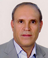
Javad Faryabi has graduated from dentistry school of Shiraz University of Medical Sciences in IRAN (1990) and postdoctoral studies from Shahid Beheshti University of Medical sciences in Tehran, IRAN(2000) . He is the director of residency program of maxillofacial surgery in Kerman University,and head of Oral & Maxillofacial deprtment of this university. He has published more than 20 papers in reputed journals and has been serving as an editorial board member of JOHK inthe mentioned above unhversity.
Abstract:
Currently, when a dentist seeks a treatment for facial swelling of his/her patients, the first diagnosis and cause of swelling that he/shet thinks about it, may be and perhaps always is the teeth of the patients and an finally is an odontogenic infection. But sometimes it is not true, and this is not the correct answer to the patients for managing their discomfort, because they are worrying about their health, and may go to several offices of dentists or other physicians and specialists for treatment of their disease, actually they need to additional work ups like CT-Scans, CBCT, MRI, Sonography, and etc.to discover the exact cause of their facial swelling, so In my presentation I will introduce to several example of such facial swelling in my patients related to maxillofacial region and proper management of them.
Gianni Frisardi
Orofacial Pain Centre, Italy
Title: NGF-Neuro Gnathological Functions: A new Trigeminal Electrophysiological Paradigm in masticatory rehabilitation
Time : 11:40-12:00
Biography:
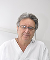
Gianni Frisardi has completed his bachelor’s degree in Medicine and Dentistry in 1983 and 1987 respectively at the “La Sapienza†University in Rome. In 1993 he attained a Master in Bioengineering and his experimental thesis “Mandibular Kinematics has been awarded by the ENEA Institute. He worked for several years in the trigeminal neurophysiology laboratory in order to transfer the know how in the field of the gnathology. He published papers in reputed journals and in 2014 was candidated as Editor by the World Dental Federation for the International Dental Journal. He has been Visiting Professor at the University of Rome, Chieti, Ancona and currently at the University of Sassari in the Department of Masticatory Rehabilitation and Orofacial Pain.
Abstract:
The masticatory system should be considered a "Complex System", which is divided into a number of constituent elements bound toghether in a set of non-linear and stochastic relationships. The result of all the constituent elements determines an "Emergent Behaviour" of the system itself. The NGF Paradigm puts the excitability of the central and peripheral masticatory pathways and the network of brain connections at the centre of this complex system. To achieve this purpose it has been necessary to create a bridge between Neurophysiological Trigeminal knowledge technologies and those of Gnathology, hence the term "Neuro Gnathological Functions". Neurophysiological procedures aim to establish the presence or absence of the integrity of the neuromuscular system through the motor evoked potentials of the bilateral trigeminal roots (bRoot-MEPs) using electrical and/or magnetic transcranial stimulation methods (eTCS and mTCS). This first procedure would be able to generate a “Normalization Factor†to which to refer all trigeminal reflexes, bypassing the maximum voluntary contraction (MVC), which is too variable and unstable. In prosthetic implant rehabilitation and in patients with TMDs, however, these neurophysiological trigeminal procedures are capable of determining an intermaxillary spatial relation called “Neural Evoked Centric Relationâ€. Further, the study of trigeminal reflexes, normalized to the bRoot-MEPs represent a potential clinical support in masticatory rehabilitation procedures such as orthodontic treatment, implant prosthesis and in the differential diagnosis of Orofacial pain. Some clinical cases will be presented in order to better identify the clinical support that this paradigm could add to the dental sciences.
Flavio Frisardi
Orofacial Pain Centre, Italy
Title: NGF- Trigeminal electrophysiology in complex orthodontic treatements
Time : 12:00-12:20
Biography:
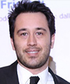
Degree in Orthodontics and Prosthetic Dentistry obtained at the Polytechnic University of Ancona on 22/10/2003, with a thesis on “Trigeminal electrophysiology in patients with Orofacial pain and Temporomandibular disorders†and he published correlated articles on reputed International journals. For this reason, he has joined to the NGF research field in order to transfer the trigeminal electrophysiological technologies into the orthodontic treatments, adding scientific and professional value to the discipline.
Abstract:
In some cases, the patients who suffer temporomandibular joint or orofacial pain complains experience an improvement following the orthodontic treatment, while in other circumstances at the end the treatment the patient develops an occlusal dysfunction, which may be aggravated by dislocations of the meniscus and muscle pain. The Neuro Gnathological Functional technology (NGF) now makes it possible for us to monitor the progress of the orthodontic treatment and identify any irritative spine introduced in the system. For instance, in the cases for which an occlusal vertical increasing is required, the symmetry of the jaw reflexes is of major importance, since the unlikely event of occlusal asymmetry could deteriorate causing a more pronounced malocclusion at the end of the treatment. In all other cases of dental crowding, even without modifications in the vertical dimension, the trigeminal electrophysiological reflexes should be monitored so as not to lose the occlusal centricity and change the neuromuscular bio-mechanics laws. This result would give added value target for the efficiency of orthodontic treatment and patient safety. Complex clinical cases will be discussed, for which the functional neuro gnathological support has been decisive in the achievement of the set target.
Mohammad Dib Kanaa
Newcastle University,UK
Title: Efficacy of dental local anaesthesia in mandibular teeth: Current views
Time : 12:20-12:40

Biography:
Dr Mohammad Dib Kanaa is currently working as a specialty Doctor in Oral and Maxillofacial Surgery at Kettering General Hospital NHS Foundation Trust. Dr Kanaa also worked at different British Universities and National Health Service Hospitals. He has worked as a Doctor in Oral and Maxillofacial Surgery Departments at South Yorkshire (Rotherham General District Hospital, Doncaster Royal Infirmary, Mexbrugh and Montague). Most recently he has worked at Southend University Hospital in the South East of the United Kingdom as a Doctor in Oral and Maxillofacial Surgery Department that included four main hospitals in Essex (Southend University Hospital, Basildon, Colchester and Broomfield Hospitals). In 2013, he was involved in the Dental Treatment and Oral and Maxillofacial Surgery Management at Galeazzi Hospital, Milan Italy. He has a degree in Dentistry (DDS) as well as a degree in the History of Medical Sciences. Dr Mohammad Kanaa was awarded his Master degree in Oral and Maxillofacial Surgery at Manchester University, UK. Dr Kanaa has also achieved his PhD degree in Oral and Maxillofacial Surgery at Newcastle University, UK where he gained the best Endodontic Research Prize in the UK in 2006 for Efficacy of Local Dental Anaesthesia. Dr Kanaa has published significant number of key papers in peer review journals in collaboration with others British Scientists and Dentists and wrote a number of important books for dental literature as well as significant number of conference presentations nationally and internationally.
Abstract:
Objectives: The current research work evaluated the efficacy of different local anaesthetic techniques/solutions in volunteer trials and its effectual outcomes for patients suffering mandibular irreversible pulpitis teeth. Methods: Double blind randomized cross over studies were investigated in the mandibular teeth in volunteers. One clinical study included 205 patients suffering from irreversible pulpitis in the mandibular teeth. Lidocaine and articaine local anaesthetic drugs were used in these trials. Electronic pulp tester was used in all studies. No response to negative pulp tester (80 reading) was the criterion of profound teeth pulp anaesthesia. Visual Analogue Scale (VAS) was employed to report the total discomfort experience and the total experience of treatment discomfort. Results and conclusions: Slow inferior alveolar nerve block (IANB) was more effective than rapid IANB injection for mandibular first molar, premolar and later incisor pulp anaesthesia. Slow injection was more comfortable than rapid injection following lidocaine IANB and incisive mental nerve block injections. Infiltration anaesthesia with articaine was more effective than lidocaine for mandibular first molar pulp anaesthesia. Articaine buccal infiltration was as effective as IANB injection for lower first molar pulp anaesthesia. Articaine buccal infiltration supplemented to lidocaine IANB was more effective than lidocaine IANB injection alone. Infiltration anaesthesia was more comfortable than IANB injection. Approximately 45% of mandibular teeth experienced successful treatment following lidocaine IANB. Articaine buccal infiltration and intraosseous injection techniques were superior to intraligamentary and repeat IANB injections following failure of IANB injection. Injections and treatment discomfort were in the mild category.
Mohamed Khaled Ahmed Azzam
King Abdulaziz Medical City -National Guard Hospital, Saudi Arabia
Title: Nociceptive Trigeminal Inhibition Reflex (NTI) Tension suppression system (TSS)
Time : 12:40-13:00
Biography:
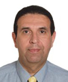
Dr Azzam graduated from Cairo University in 1976 followed by a training program from Minnesota Dental School-Minnesota-USA in Surgery in 1982. He finished his Master’s degree-M.D.Sc in 1984 in Removable Prosthodontics from Cairo University in 1984 and then completed one year fellowship in Removable Prosthodontics in Tufts dental School, Boston –Massachusetts –USA 1989-1990. He finished his PhD –D.D.Sc in Removable Prosthodontics 1993 after which was teaching in King Abdulaziz Dental School in Saudi Arabia for ten years as an Assistant Professor in Removable Prosthodontics. Currently he is the Removable Prosthodontic Consultant in King Abdulaziz Medical City – National Guard Hospital – WR, Saudi Arabia He presented over 100 National and International lectures all over the world and in 2009 was awarded Best Oral presentation in 31st Asia Pacific Dental Congress.
Abstract:
Tension headache patients without symptoms of Temporal-mandibular disorders (TMD) contract their temporalis muscles during sleep 14 times more intense than upon awakening. Once the jaw is clenched, the supportive musculature of the skull assumes a static contraction causing chronic stiff and sore neck. Night guards could relax the lateral pterygoid musculature by providing less resistance to side-to-side movement. When the lateral pterygoids are chronically contracted (dysfunctional habit) they can cause sinus symptoms and /or TMD .If the patient’s parafunctional habits includes both horizontal activity ( lateral pterygoid) and vertical activity ( temporalis) the result is “excursive clenching or bruxing†which allows significant TMJ strain. But if the parafunctional habit is purely vertical (temporalis) the result is “primary clenching†which causes severe morning headaches.
During this unprotected nocturnal parafunction, massive amounts of noxious input called nociception bombard the trigeminal sensory nucleus. By keeping the molars and canines from touching the method of generating “Nociception to the Trigeminal†is inhibited.
In conclusion an inter-occlusal device which provides anterior incisor contact only will reduce the contraction intensity of the temporalis significantly. This occurs only in a static position, so modifications of the device to avoid lower canine contacts during excursive movements are done as:
• Constructing an Anterior Midline Point Stop (AMPS) will avoid canine contact to the device during lateral movements hence Headaches and TMD are prevented.
• Further modification to maintain perpendicular incisal contact in protrusive and retrusive movements allows for suppression of parafunctional contraction intensity in all mandibular movements thereby reducing and preventing Headaches and TMD symptoms.
Lunch Break 13:00-13:45 @ Al Tanour Restaurant
Marwa Sharaan
Gulf Medical University, UAE
Title: Microbiological analysis of teeth with chronic apical periodontitis and the outcome of treatment by gutta percha points impregnated with either calcium hydroxide or chlorhexidine as intra canal medicament
Time : 13:45-14:05
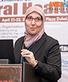
Biography:
She is an Assistant Professor of Endodontics at GMU, Ajman, UAE. She was a lecturer at the Department of Endodontics Faculty of Dentistry, Suez Canal University, Egypt. She got her Bachelor of Oral Surgery & Dental Medicine (BDS), Faculty of Dentistry, Alexandria University, Egypt in 1996. In 2003, she had her Master Degree in Conservative Dentistry (Endodontics) (MSc), Faculty of Dentistry, Suez Canal University, Egypt. Subsequently, she got her Doctoral Degree in Dental Sciences (Endodontics) (DDSc), Faculty of Dentistry, and Suez Canal University, Egypt in 2009. She is a member of many of the dental associations. She has several publications. She shared in various conferences as a speaker as well as a chairperson.
Abstract:
Bacteria have a major role in the success of an endodontic treatment. Mechanical preparation alone cannot effectively eliminate bacteria from dentinal tubules and other irregularities in the root canal. Few remnant micro‑organisms can multiply between appointments often reaching the same level as at the start of the previous session. These observations call for an effective intra‑canal medication that will help to disinfect the root canal system. Calcium hydroxide has an antibacterial effect on most of the endodontic pathogens. This is due to its high alkalinity. It damages the bacterial cytoplasmic membrane. Chlorhexidine has been found to have a broad-spectrum antimicrobial action and relative absence of toxicity. Calcium hydroxide points as well as chlorhexidine gutta percha points are ready to be used in a flexible way and easily to be introduced into the canal. Three bacterial cultures - before, after instrumentation and after medication by either one of the experimental materials - were taken from forty healthy patients having non-vital single rooted teeth with chronic apical periodontitis. Twelve types of bacteria were commonly isolated with different frequencies. Anaerobes represented about 56.1 % and aerobes represented about 43.9 % of the total bacteria. Streptococcus mitis (15.2 %) was the most frequent isolated bacteria (aerobes and anaerobes) from all the cases in the 1st culture. Then it was followed by Prevotella intermedia (14.7 %), Peptostreptococcus spp. (10.5%), Eubacterium lentum (9.4 %), Lactobacillus spp. (8.9%), Staphylococcus aureus (8.4 %), Veillonella parvula (7.9 %), Enterococcus faecalis (7.3 %), Escherichia coli (E-coli) (6.8 %), Neisseria catarrhalis (5.2%), Proionibacterium spp. (4.7 %), Staphylococcus epidermis (1 %). The results demonstrated a highly significant reduction in the bacterial growth in the experimental groups before and after instrumentation and medication of the root canal when compared to the control. Chlorhexidine gutta percha points proved to have a significant better antibacterial effect than calcium hydroxide gutta percha points.
Jules Poukens
Cranio-maxillofacial Surgeon Atrium/Orbis Medical Center, The Netherlands
Title: NEED A NEW SKULL OR MANDIBLE? 3D PRINT IT !
Time : 14:05-14:25
Biography:
Prof. Dr. Jules Poukens is currently lecturer and researcher at the Biomed Research Institute of the University Hasselt and the University of Leuven in Belgium. He is a Cranio-Maxillofacial Surgeon at the Medical Center Sittard /Heerlen in the Netherlands. He got his M.D. and D.M.D from the Catholic University Leuven in Belgium. He was trained as Cranio-Maxillofacial Surgeon in Belgium (Leuven) and Germany (Freiburg, Black Forrest). After his training, he first joined the staff at the University Hospital Maastricht and later on the staff of Medical Center Sittard /Heerlen in the Netherlands. He has participated in several European Community funded projects on medical rapid prototyping and manufacturing and was acting chairman of the Board of Directors of the EU funded project CUSTOM-IMD. He was the leading surgeon that designed and implanted the world’s first 3D printed total mandibular implant and was one of the pioneers in using 3D printed skull implants. He has numerous publications and held numerous presentations in this field. Born and raised in Belgium, Jules searched for his roots and lives with his wife and two daughters in Dilsen, Belgium near the Dutch and German border.
Abstract:
Patients in the cranio-maxillofacial clinic often present with serious, complex, and potentially life- threatening or life-limiting medical conditions (e.g. tumor, trauma, aggressive osteomyelitis). Available treatments may not always give satisfactory results for patients and doctors. Therefore, complex problems ask for new solutions. An emerging technique in the medical field is Computer Aided Design (CAD) , Computer Aided Manufacturing (CAM) by 3D printing. For successful implementation of CAD-CAM technology in the clinical practice doctors, dentists and engineers need to work together and share their expertise. This intense cooperation leads to 3D printing of custom patient specific implants. 3D printed implants are used for the treatment of skull defects, dental superstructures and world’s first 3D printed entire mandible replacement implant. These clinical cases will be highlighted.
Roula Al Bounni
Riyadh Colleges of Dentistry & Pharmacy, Kingdom of Saudi Arabia
Title: Direct composite veneers as a permanent treatment of anterior teeth
Time : 14:25-14:45
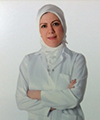
Biography:
Roula AL-Bounni currently is professor in operative dentistry. She has worked with undergraduate and postgraduate students in clinical & literature aspects in Operative & Endodontic department 1995-2011. She supervised more than 15 MSc and 6 PhD student’s certificates in Operative and aesthetic dentistry 2002 -2011. She was Head of Endodontic & Operative Dentistry Department 2007-2011
Abstract:
Every new material or technique introduced to the field of dentistry aims to achieve esthetic and successful dental treatments with minimal invasiveness. Therefore, direct laminate veneers have developed for advanced esthetic problems of anterior teeth. Tooth discolorations, rotated teeth, coronal fractures, congenital or acquired malformations, diastemas, discolored restorations, palatally positioned teeth, abrasions and erosions are the main esthetic problems for many patients, and it's basically the main indications for direct laminate veneers. Also, along passing time, Changes in color, shape, and structural abnormalities of anterior teeth might lead to important esthetic problems for patient, for that laminate direct composite veneers could be a good conservative solution for all these problems. Direct Laminate veneers are applied on prepared tooth surfaces with composite resin material directly in dental clinic. Absence of necessity for tooth preparation, low cost, intraoral polishing of direct laminate veneers is easy to use, and any cracks or fractures on the restorations may be repaired intra-orally, all of these are some advantages of this technique, however, the main disadvantages of direct veneers are low resistance to wear, discoloration and fractures, but the improvements of the new generations of resin composite restorations and adhesive techniques have surmount all these problems. But it's worth mentioning that the material itself, and the ideal application of this technique is the main factor to achieve long lifespan for successful outcome. By this technique we can get lot of benefits with wide range of people who seek improvements in there distorted smile due to numerous problems in anterior teeth.
Wisam Al-Rawi
University of Detroit Mercy School of Dentistry, USA
Title: The current state of Cone Beam CT (CBCT) 3D imaging in dentistry
Time : 14:45-15:05
Biography:
Wisam Al-Rawi has completed his MSc degree in medical imaging (oral imaging option) from K.U.Leuven, Leuven, Belgium in 2006 and MS degree in oral and maxillofacial radiology from the University of North Carolina at Chapel Hill, NC, United States in 2009. He is currently holding an adjunct faculty position at the University of Detroit Mercy School of Dentistry and studying in the Accelerated Dental Program. He founded Marcilan Inc. A company dedicated for oral radiology education and created two educational apps in oral radiology for iPhone and iPad: CBCT and iPanoramic. He helped two dental schools to move to digital radiography. He has published several papers regarding Cone Beam CT and oral radiology education.
Abstract:
The introduction of Cone Beam CT (CBCT) constitutes a paradigm shift in the way clinicians acquire and view radiographs. Unlike intraoral or panoramic radiographs, there is no magnification and no superimposition of structures. CBCT volumetric imaging provides unprecedented highly detailed radiographic information about the patient’s teeth, bone, jaws, and airways spaces. This information can be used for diagnosis and treatment planning including implant planning, detection of root fractures, assessing the relationship of an impacted wisdom tooth to the mandibular canal, sleep apnea studies, growth assessment, pre and post-surgery assessment, and pathology. However, with CBCT, there is increased financial costs and radiation exposure to the patient. It is important that clinicians understand the advantages and limitation of this technology for better diagnosis and treatment planning of their patients. With many machines available on the market and more coming each year, it can clearly be seen that this technology is taking a big role in the field of dentistry. This purpose of this presentation is to: (i) review principles of action of CBCT; (ii) compare radiation dose of different machines; (iii) highlight clinical cases for use of CBCT in dentistry and how it can be used as a tool to aid in diagnosis and treatment planning of different cases in dentistry.
Carolina Duarte
Ras Al Khaimah College of Dental Science, UAE
Title: The potential of drug and gene therapy applied to orthodontic treatment
Time : 15:05-15:25

Biography:
Dr. Duarte did her pre-graduate training at the National Autonomous University of Honduras and obtained her PhD in Maxillofacial Orthognathics from Tokyo Medical and Dental University in Japan. She has studied the effect of hormones on bone metabolism and their application in orthognathic treatment, and presented and published her research internationally. She recently joined the faculty at Ras Al Khaimah College of Dental Science.
Abstract:
Interceptive and comprehensive orthodontic treatments are characterized by the use of appliances that mechanically modify bone size and shape, and reposition teeth in the dental arches. The bone modifications and tooth movements done with these orthodontic appliances can be complex and painful, require long retention time and are subject to significant amounts of relapse. Orthodontic research has focused on optimizing the mechanics, materials and esthetics of orthodontic appliances which have made orthodontic treatment more predictable and acceptable to patients. However, less importance is given to therapeutic adjuvants that may optimize bone remodeling during orthodontic treatment and improve hard tissue adaptation after treatment is completed. Numerous reports have been published on the effects of drugs or hormone therapy during orthodontic treatment, as well as the genes involved in bone remodeling during orthodontic tooth movement. This basic knowledge could be translated to safe clinical therapies with the potential to reduce treatment and retention time, facilitate anchorage and control relapse. Gene therapy is trending in many areas of medical and dental practice but has been overlooked by orthodontists. It is only logical to wonder if orthodontic treatment will continue to grow as a purely mechanic therapy or will evolve to a hybrid practice using mechanics and gene therapy to optimize its progression and results.
Lamis Abuhaloob
Ministry of Health, State of Palestine
Title: School dental health in Palestine
Time : 15:25-15:45

Biography:
Lamis abuhaloob obtained PhD in Dental Public Health from Newcastle University in the United Kingdom. She is Head of Oral and Dental School Health in Ministry of Health in Palestine. She has published and presented several papers in national and international journals and conferences of dentistry.
Abstract:
By the end of 2012, Palestinians (including refugees) living in Palestinian territories were 5.8 million, approximately, 40% of them were school children. Since the eve of the 1948 war, the Occupied Palestinian Territories have suffered from political conflict, resulted in serious deteriorations in the socio-economic status and psycho-social wellbeing of the Palestinian people. World Health Organization stressed the high burden of oral diseases and its adverse effect on general health and quality of life. Several studies found that oral diseases have profound impact on children’s performance in schools, nutrients intake, and growth. Despite, school dental health programme in Palestine have been established to promote general and oral health among school children in government and UNRWA schools, there was a gradual increase in the prevalence of dental disorders and mainly dental caries. Generally, the def was higher than DMF. Incidences of periodontitis, fluorosis and malocclusion were the highest in the year of Al-Quds Intifada upraise, and in 10th grade schoolchildren. Increased oral and dental diseases in children in Palestine have overloaded the health services and caused a negative impact on the effectiveness of prevention programs and health promotion strategies in both of Palestinian Ministry of Health and UNRWA. Political unrest had a profound negative effect on oral health in Palestine. The increase in rate of decayed teeth in schoolchildren is alarm and suggests an increase in risk of dental caries in future generations. School dental health programme struggles to improve child oral and dental health. Thus, It should be revised. Effective oral health promotion interventions should consider age specific oral diseases and political unrest.
Coffee Break 15:45-16:00 @ Al Diyafah Pre-function area
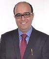
Meheriar Chopra
Dr Meheriar’s Dental Clinic, India
Title: Lasers in root canal sterilization: A clinical case presentation
Time : 16:00-16:20
Biography:
Meheriar Chopra has completed his BDS and MDS in Conservative Dentistry and Endodontics from Maharashtra University of Health Sciences Ministry of Health, M.A.R Dental College, Pune, India. He has completed his Diploma in Lasers from SOLA (Society of Oral Laser Applications) affiliated to University of Vienna. He is a leading Endodontist with a private practice in Pune, India. He has research articles published in both national and international journals.
Abstract:
Lasers (Light amplification by stimulated emission of radiation) are slowly but surely revolutionizing dental treatment. In dentistry, lasers are accurate, preserve more healthy tooth structure, produce minimum to no pain during treatment, sterilize tissues with minimal to no bleeding and promote faster tissue healing. The first use of lasers in endodontics was in 1971. With a rapid development of laser technology, new lasers with a wide range of characteristics are now available. In endodontics they are used for a variety of procedures such as alleviating dentinal hypersensitivity, pulpal diagnosis, pulp capping and pulpotomy, cleaning and shaping of root canal systems, canal sterilization and endodontic surgery. The following clinical cases demonstrate the use of a diode laser in canal sterilization, promoting faster periapical healing. Studies have shown that lasers have a bactericidal effect. The laser beam as it diverges has a higher penetration depth enabling irradiation of the curvatures and complexities within the root canal system like lateral and accessory canals, fins, webs and apical deltas. Laser irradiation has the ability to remove debris and the smear layer from the root canal walls following biomechanical instrumentation. This enables the clinician to confidently clean and shape the root canal system in minimum time leading to higher patient satisfaction.
Vivek Padmanabhan
Ras Al Khaimah College of Dental Sciences Ras Al Khaimah, UAE
Title: Stress Estimation in Pediatric Dentistry – A Correlation Study between Modified Dental Anxiety Scale and Salivary Cortisol Levels
Time : 16:20-16:40

Biography:
Vivek Padmanabhan, B.D.S., M.D.S., (PhD), Assistant Professor, Department of Pediatric Dentistry, Ras Al Khaimah College of Dental Sciences, Ras Al Khaimah University of Health Sciences, Ras Al Khaimah, UAE; Tel: +971527400018; e-mail: drvivekpadmanabhan@gmail.com 1 Department of Pediatric Dentistry, Ras Al Khaimah College of Dental Sciences, Ras Al Khaimah University of Health Sciences, Ras Al Khaimah, UAE. 2 Department of Pediatric Dentistry, Bangalore Institute of Dental Sciences, Bangalore, India. 3 Department of Pediatric Dentistry, A.B.Shetty Memorial Institute of Dental Sciences, Mangalore, India
Abstract:
Anxiety is a frequent problem among dental patients in general and especially children. The presence of dental anxiety in pediatric patients is not a dilemma for the patients alone but also for the dental professionals themselves; and sometimes it renders the treatment more complicated and tedious to be accomplished successfully. Fear and anxiety increase the activity of the Hypothalamic Pituitary Axis (HPA) which in turn enhances secretion of cortisol. Cortisol also known as the stress hormone, is secreted from the adrenal cortex and dispersed to all body fluids, and can be detected in urine, serum or saliva. Heightened cortisol levels are thus indicative of increased stress as a result of elated fear and anxiety. Anxiety scales have been used for the assessment of anxiety in children. Most successfully used anxiety scale is the modified dental anxiety scale (MDAS).
120 children who required dental procedures were included in the study of which 60 each belonged to the study and control groups. For each of these children saliva was collected to evaluate salivary cortisol levels and MDAS was duly filled with the help of the parents to evaluate the anxiety levels.
The results indicated that the salivary cortisol levels and anxiety levels were significantly increased in the study group when compared to the control group. A positive correlation can also be seen between the two measurement methods of anxiety levels.
Keywords: Anxiety, Stress, Cortisol, MDAS
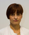
Jadranka Handzic
University hospital center, Croatia
Title: Tympanometric findings in cleft palate patients: influence of age and cleft type
Time : 16:40-17:00
Biography:
Jadranka Handžić graduated in 1984y. at Medical School University of Zagreb, took Master degree at 1987y. And PhD at 1989. Since 1987y she spent her residential program of Otolaryngology than diploma at 1989 as specialist of Otolaryngology and 2003y as sub-specialist of Audiology. At 2000-2001 she spent academic year on Fulbright Scholarship at Cleft Palate-Craniofacial Centre and Dental School of Medicine, University of Pittsburgh and Children’s Hospital Pittsburgh U.S.A Department for Paediatric Otolaryngology, University of Pittsburgh, U.S.A at position as Adjunct Associate Professor of Oral Medicine and Pathology and at 2001-2002 as Visiting Assistant Professor at Department of Oral Medicine and Pathology, Cleft Palate-Craniofacial Centre, Dental School of Medicine, University of Pittsburgh, U.S.A. 2001-2002 she had Lester Hamburg- Research Fellowship in Department for Paediatric Otolaryngology Children's Hospital of Pittsburgh, Medical School University of Pittsburgh, U.S.A. From 2002 she was Assistant Professor of Otolaryngology, University Clinical Hospital Centre and Medical School “Zagreb†and from 2008 Professor of Otolaryngology and Audiology. She is author and co-author of 16 articles in Current Contents, lecturer of 30 presentations on International conferences, reviewer in Journal and Annals of Maxillofacial Surgery, Director of 7 post-graduate studies of Otolaryngology and Audiology, author and co-author of 4 books, author of 3 international projects.
Abstract:
Objective: Conductive hearing loss associated with otitis media with effusion is usually finding in cleft lip and palate children. Method: Tympanometry performed in 57 children with bilateral (BCLP), 122 unilateral cleft lip and palate (UCLP) and 60 children with isolated cleft palate (ICP) according to age sub-groups, median age of groups 5y. All children undergo no previous ear surgery and undergo standard method of cheilognathopalatoplasty. Results: B type is most frequent in ICP (58.3%) less in UCLP (50.6%) and lowest in BCLP (46.5%).UCLP children show higher decrease B type ears with aging (rs=-0.4430) than BCLP (rs=-0.3186) and ICP (rs=-0.3378). Moderate hearing ears (21-40dB) showed highest decrease of B type with aging, lower decrease (rs=0.2184) have ears with mid threshold 11-20dB and lowest showed ears with severe hearing loss. At age 1-3y UCLP have higher rate of B type ears than BCLP and ICP which start to decrease at age 7-12y.At BCLP B type ears increase in frequency at age 4-6y.In ICP patients decrease in B type is not significant until 15y when first decrease showed. Type A is typical for older ages while B type for younger ages with significant decrease at age 4-67 and 7-9y no ear side difference. BCLP showed increase at 4-6y and the slowest decrease than other types. Conclusion: Cleft types due to craniofacial characteristics with aging have different mode of pathophysiological changes of middle ear desease.
Abeer Abo El Naga
King Abdulaziz University, Saudi Arabia
Title: Dentinal hypersensitivity: recent trends in the treatment
Time : 17:00-17:20
Biography:
Associate Professor and course director of third year Operative Dentistry at Faculty of Dentistry, king Abdulaziz University. Received my B.Sc. of Oral and Dental Medicine (1988), MSc in Restorative Dentistry, and PhD in Operative Dentistry (2007) from Faculty of Oral and Dental Medicine, Cairo University. I Served as Assistant Professor of Operative Dentistry at Misr University for Science and Technology (Cairo, Egypt) and Ibn Sina National College for Medical Studies (Jeddah, Saudi Arabia) from 2007 to 2011. I had presented and published more than 20 papers in reputed journals.
Abstract:
Dentine hypersensitivity is one of the most encountered problems in the dental clinics. When a patient presents with dentine hypersensitivity symptoms, he/she should be examined and informed of the proper treatment option to eliminate the problem. However, many dentists have some problems in diagnosing and treating dentine hypersensitivity. Selection of the most appropriate treatment option to achieve successful management is based on proper diagnosis for the condition and its etiological factor. This presentation will cover the following: the etiological factors, diagnosis and invasive vs. most recent non invasive treatment of dentine hypersensitivity.
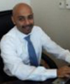
Mohammad Abdulwahab
Kuwait University, Kuwait
Title: The Need for Anesthesia and Sedation Services in Kuwaiti Dental Practice
Time : 17:20-17:40
Biography:
Mohammad abdulwahab has completed his dental anesthesiology and publich health training from University of Pittsburhg and postdoctoral studies in managment of special needs dental patients from the same institute. He served as a faculty if univeristy of pittsbugrh school of dental medicine. Currently he is a faculty member at kuwait university faculty of dentistry. He a member in number of comittes and a mentor for residents. His research foucus on medical emergency and pain control. He has published in reputed journals and has been serving as consultante for training.
Abstract:
The public health relevance of the prevalence of dental fear in Kuwait and the resultant barrier that it creates regarding access to dental care was assesded. An analysis demonstrated a high prevalence of dental fear and anxiety in the Kuwaiti population and a perceived need for anesthesia services by dental care providers. The assessment of the general population showed nearly 35% of respondents reported being somewhat nervous, very nervous, or terrified about going to the dentist. In addition, about 36% of the population postponed their dental treatment because of fear. Respondents showed a preference to receive sedation and anesthesia services as a means of anxiety relief, and they were willing to go to the dentist more often when such services were available. People with high fear and anxiety preferred to receive some type of medication to relieve their anxiety. In conclusion, the significance and importance of the need for anesthesia services to enhance the public health of dental patients in Kuwait has been demonstrated, and improvements are needed in anesthesia and sedation training of Kuwaiti dental care providers.
Mustafa DAG
Gulhane Military Medicine Academy, Turkey
Title: The Maxillary Sinus Membrane Elevation Technique Using Venous Blood Without Grafting Material And Immediate Implant Placements: A Case Series Analysis
Time : 17:40-18:00
Biography:
Mustafa DAÄž graduated from Ankara University Faculty of Dentistry in 2007. He began his doctoral studies at Oral and Maxillofacial Department of Gulhane Military Medicine Academy in 2011 and is still ongoing. He is working especially on implant placement and bone augmentation topics. He has published more than 5 papers in national or international journals. He participated in many national and international congresses with oral or poster presentations.
Abstract:
Introduction: Various sinus augmentation techniques have been successfully used in order to enable the implant placements in the atrophic posterior maxilla. The object of the study was to evaluate whether sinus membrane elevation technique using venous blood without any grafting material constitute an effective technique for the maxillary sinus augmentation. Method: Eighteen patients who have not sufficient bone for implant placement at maxillary posterior sides were included in the study. Twenty-eight dental implants were inserted to twenty-two maxillary sinus just after the sinus membrane elevations via lateral approaches. After implant placements the cavity between relocated schneiderian membrane and sinus inferior wall was filled with venous blood without any bone grafting material. Cone beam computerized tomography was used for follow-up evaluation. Results: Comparisons of pre- and postoperative radiographs clearly demonstrated new bone formation between the new and former sinus inferior walls. Within the mean follow-up period of 1.5 years two implant failures were recorded. One of the failures was occurred just three months after the surgery due to infection, and the other implant failure was recorded almost a year after the prosthetic rehabilitation. The survival rate was nearly % 93 in the study. In a systematic review by Wallace et al, it was reported that average survival rate of implants placed in maxillary sinuses augmented with the lateral window technique was nearly %92. It was shown that the technique used in this study has provided high implant survival rate as studies using grafting materials for maxillary sinus augmentation. Conclusion: The case series of 18 patient demonstrated that the maxillary sinus membrane elevation procedure without grafting material and immediate implant placements is considered to be more cost-effective and less time-consuming compared with conventional procedure.
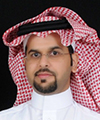
Sultan Aldeyab
Ministry of National Gauard, King Abdulaziz Medical City, Riyadh
Title: Correction of Excessive Spaces in the Esthetic Zone
Time : 18:00-18:20
Biography:
Sultan Aldeyab has completed his AEGD certificate with honor in 2008, and then he got Saudi Board of Restorative Dentistry with honor from 2008-2012. He is working in Restorative Department in King Abduaziz Medical City (National Guard) Riyadh, He is teaching restorative post graduate resident and lecturing undergraduate student in dental collage of King Saud Bin Abdulaziz University for Health Sciences. He got many awards and he has many publications and lectures.
Abstract:
Introduction:
The use of porcelain crowns and veneers to solve esthetic problems has been shown to be a valid management option especially in the anterior esthetic zone. This case report discusses a patient having diastema in the anterior region. The patient was treated with orthodontic treatment and porcelain crowns & veneers in the maxillary arch for the closure of diastema.
Clinical report:
A 35 year old male patient with a chief complaint of discolored anterior teeth and gaps between the teeth. The patient was unhappy with the appearance of his teeth.After thorough examination; impressions for diagnostic models were made in irreversible hydrocolloid. The models were studied to decide the shape and size of the restorations with help of a diagnostic wax up. Before proceeding for tooth preparation, shade was selected using Classical shade guide (Vita Zahnfabrik, Germany). The maxillary teeth were then prepared from right 2nd premolar to the left 2nd premolar to receive porcelain crowns and laminate veneers. Impression of the maxillary arch was made in addition silicone.
The laminates were etched with 4 % Hydrofluoric acid, after etching, they were washed thoroughly using liberal amount of water. On drying, a coat of Silane coupling agent (Porcelain Primer, Bisco,USA ) performed on two teeth at a time starting at the midline.
The prepared teeth were etched using 37% Phosphoric Acid for 15 seconds. On air drying bonding agent was applied & light cured for 10 seconds. Composite luting agent was used for cementation. The laminates were spot cured for 5 seconds initially. Excess cement was removed with explorer and then complete curing was done for 20 seconds. On completion of the cementation procedure, the occlusion was checked in centric and eccentric positions for interferences. The high points were removed and polished.
Discussion:
The etiology of diastema may be attributed to the following factors: (a) Hereditary- congenitally missing teeth, tooth and jaw size discrepancy, supernumerary teeth & frenum attachments; (b) Developmental problems- habits, periodontal disease, tooth loss, posterior bite collapse (Oesterle & Shellhart, 1999). Treatment planning for diastema correction includes orthodontic closure, restorative therapy, surgical correction or multidisciplinary approach depending upon the cause of diastema (Dlugokinski et al, 2002).
Conclusion:
Orthodontic treatment, bonded porcelain crowns & veneers can provide successful esthetic and functional long-term service for patients.
- Advanced Course

Chair
Fabio Savastano
Preseident, ICNOG, Italy
Session Introduction
Fabio Savastano
International College of Neuromuscular Orthodontics and Gnathology, Italy
Title: Neuromuscular orthodontics: The future is here
Biography:
Fabio Savastano is a Medical Doctor from Italy with over 25 years of experience in Orthodontics and Gnathology. He did Postgraduate Master in Orthodontics from the University of Padua, Italy, founded Neuromuscularorthodontics.com (now ICNOG) in 1994. He has lectured in India, Middle East, Canada, Brazil and Italy and he is President of the International College of Neuromuscular Orthodontics and Gnathology (ICNOG). He practices in Albenga, Italy, and is limited to orthodontics and TMD.
Abstract:
The key in understanding occlusion and malocclusion is the analysis of function. Neuromuscular Orthodontics is a relatively new and easy approach engineered for the orthodontist of the future. This brief course has the objective to introduce you to the basics of Neuromuscular Orthodontics: Muscle function (EMG), lip balance, cranio-cervical posture, mandibular tracking, muscle relaxation (T.E.N.S.). During this course you will be shown several cases treated with a variety of functional appliances for the treatment of Class I-II-III malocclusions and oral orthotics for the treatment of TMJ disorders. See how easy treatment is once your comprehension of the cause of malocclusion is exhaustive. Part 1 of the course will focus on how to diagnose a malocclusion without all the complicated tasks that have been taught in the past, but in a more modern and functional fashion. Part 2 will show you how to build a better looking face while reducing relapse and TMJ problems.
Why should they attend?
1. This course will open your mind as an orthodontist. Understanding the hidden functional characteristics of the individual is a must in planning an ideal treatment.
2. Where is the mandible supposed to be relative to the cranium? The correct cranio-mandibular relationship guarantees an optimal TMJ function: A key to success.
3. See mandibular advancement in growing and adult patients: Build beautiful faces.
